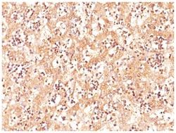
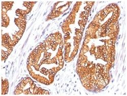
TYRP1 Mouse, Clone: SPM456, Novus Biologicals™
Mouse Monoclonal Antibody
Manufacturer: Fischer Scientific
The price for this product is unavailable. Please request a quote
Antigen
TYRP1
Dilution
Flow Cytometry 0.5-1ug/million cells, Immunocytochemistry/Immunofluorescence 1-2ug/ml, Immunohistochemistry-Frozen 0.5-1ug/ml
Classification
Monoclonal
Form
Purified
Regulatory Status
RUO
Formulation
PBS with 0.05% BSA. with 0.05% Sodium Azide
Gene Alias
b-PROTEIN, CAS25,6-dihydroxyindole-2-carboxylic acid oxidase, Catalase B, CATB, EC 1.14.18, EC 1.14.18.-, EC 1.14.18.1, Glycoprotein 75, GP75DHICA oxidase, Melanoma antigen gp75, TRP-1, TRPTYRP, tyrosinase-related protein 1TRP1, TYRRP
Gene Symbols
TYRP1
Isotype
IgG2a κ
Purification Method
Protein A purified
Test Specificity
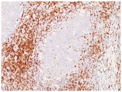
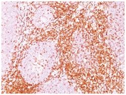
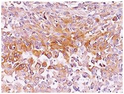
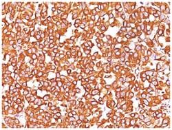
It reacts with a 75kDa melanocyte-specific gene product, identified as Tyrosinase-related protein-1 (TRP-1). It is involved in melanin synthesis. TRP1 is present on the melanosomal membranes of melanoma, normal melanocytes and nevi.Recent evidence suggests that TRP-1 is involved in maintaining stability of tyrosinase protein and modulating its catalytic activity. TRP-1 is also involved in maintenance of melanosome ultrastructure and affects melanocyte proliferation and cell death.
Clone
SPM456
Applications
Flow Cytometry, Immunocytochemistry, Immunofluorescence, Immunohistochemistry (Frozen)
Conjugate
Unconjugated
Host Species
Mouse
Target Species
Human, Mouse
Gene Accession No.
P17643
Gene ID (Entrez)
7306
Immunogen
SK-MEL-23 cells
Primary or Secondary
Primary
Content And Storage
Store at 4C.
Molecular Weight of Antigen
75 kDa
Related Products

Description
- TYRP1 Monoclonal specifically detects TYRP1 in Human, Mouse samples
- It is validated for Flow Cytometry, Immunocytochemistry/Immunofluorescence.
Compare Similar Items
Show Difference

Antigen: TYRP1
Dilution: Flow Cytometry 0.5-1ug/million cells, Immunocytochemistry/Immunofluorescence 1-2ug/ml, Immunohistochemistry-Frozen 0.5-1ug/ml
Classification: Monoclonal
Form: Purified
Regulatory Status: RUO
Formulation: PBS with 0.05% BSA. with 0.05% Sodium Azide
Gene Alias: b-PROTEIN, CAS25,6-dihydroxyindole-2-carboxylic acid oxidase, Catalase B, CATB, EC 1.14.18, EC 1.14.18.-, EC 1.14.18.1, Glycoprotein 75, GP75DHICA oxidase, Melanoma antigen gp75, TRP-1, TRPTYRP, tyrosinase-related protein 1TRP1, TYRRP
Gene Symbols: TYRP1
Isotype: IgG2a κ
Purification Method: Protein A purified
Test Specificity: It reacts with a 75kDa melanocyte-specific gene product, identified as Tyrosinase-related protein-1 (TRP-1). It is involved in melanin synthesis. TRP1 is present on the melanosomal membranes of melanoma, normal melanocytes and nevi.Recent evidence suggests that TRP-1 is involved in maintaining stability of tyrosinase protein and modulating its catalytic activity. TRP-1 is also involved in maintenance of melanosome ultrastructure and affects melanocyte proliferation and cell death.
Clone: SPM456
Applications: Flow Cytometry, Immunocytochemistry, Immunofluorescence, Immunohistochemistry (Frozen)
Conjugate: Unconjugated
Host Species: Mouse
Target Species: Human, Mouse
Gene Accession No.: P17643
Gene ID (Entrez): 7306
Immunogen: SK-MEL-23 cells
Primary or Secondary: Primary
Content And Storage: Store at 4C.
Molecular Weight of Antigen: 75 kDa
Antigen: Vimentin
Dilution: Western Blot 0.5-1ug/ml, Flow Cytometry 0.5-1ug/million cells, Immunocytochemistry/Immunofluorescence 1-2ug/ml, Immunoprecipitation 0.5-1ug/500ug protein lysate, Immunohistochemistry-Paraffin 0.5-1.0ug/ml, Immunohistochemistry-Frozen 0.5-1.0ug/ml
Classification: Monoclonal
Form: Purified
Regulatory Status: RUO
Formulation: PBS with 0.05% BSA. with 0.05% Sodium Azide
Gene Alias: FLJ36605, vimentin
Gene Symbols: VIM
Isotype: IgG1
Purification Method: Protein G purified
Test Specificity: This MAb reacts with a 58kDa protein identified as vimentin. It shows no cross-reaction with other closely related intermediate filament proteins (IFPs) such as desmin, keratin, neurofilament, and glial fibrillary acid protein.Anti-vimentin alone is of limited value as a diagnostic tool; however, when used in panels with other antibodies, it is useful for the sub-classification of a given tumor. Expression of vimentin, when used in conjunction with anti-keratin, is helpful when distinguishing melanomas from undifferentiated carcinomas and large cell lymphomas. All melanomas and Schwannomas react strongly with anti-vimentin. It labels a variety of mesenchymal cells, including melanocytes, lymphocytes, endothelial cells, and fibroblasts. Non-reactivity of anti-vimentin is often considered more useful than its positive reactivity, since there are a few tumors that do not contain vimentin, e.g. hepatoma and seminoma. Anti-vimentin is also useful as a tissue process control reagent.
Clone: SPM576
Applications: Western Blot, Flow Cytometry, Immunocytochemistry, Immunofluorescence, Immunoprecipitation, Immunohistochemistry (Paraffin)
Conjugate: Unconjugated
Host Species: Mouse
Target Species: Human, Porcine, Bovine, Canine, Chicken, Feline, Goat, Mouse (Negative), Rat (Negative)
Gene Accession No.: P08670
Gene ID (Entrez): 7431
Immunogen: Human vimentin recombinant protein
Primary or Secondary: Primary
Content And Storage: Store at 4C.
Molecular Weight of Antigen: __
Antigen: ACTH
Dilution: Western Blot 0.5-1.0ug/ml, Flow Cytometry 0.5-1ug/million cells, Immunocytochemistry/Immunofluorescence 0.5-1ug/ml, Immunoprecipitation 0.5-1ug/500ug protein lysate, Immunohistochemistry-Paraffin 0.5-1ug/ml, Immunohistochemistry-Frozen 0.5-1.0ug/ml
Classification: Monoclonal
Form: Purified
Regulatory Status: RUO
Formulation: PBS with 0.05% BSA. with 0.05% Sodium Azide
Gene Alias: ACTH, adrenocorticotropic hormone, adrenocorticotropin, alpha-melanocyte-stimulating hormone, alpha-MSH, beta-endorphin, beta-LPH, beta-melanocyte-stimulating hormone, beta-MSH, CLIP, corticotropin-like intermediary peptide, corticotropin-lipotropin, gamma-LPH, gamma-MSH, lipotropin beta, lipotropin gamma, LPH, melanotropin alpha, melanotropin beta, melanotropin gamma, met-enkephalin, MSH, NPP, POC, pro-ACTH-endorphin, proopiomelanocortin, pro-opiomelanocortin, proopiomelanocortin preproprotein
Gene Symbols: POMC
Isotype: IgG1 κ
Purification Method: Protein A purified
Test Specificity: ACTH (same as Corticotropin) is a 39 amino acid active peptide produced by the anterior pituitary. This MAb is specific to Synacthen (aa1-24 of ACTH); does not react with CLIP (aa17-39 of ACTH). POMC (pro-opiomelanocortin or corticotropin-lipotropin) is a 267 amino acid polypeptide hormone precursor that goes through extensive, tissue-specific posttranslational processing by convertases. POMC is cleaved into ten hormone chains named NPP, ACTH, alpha-MSH (Melanocyte Stimulating Hormone), beta-MSH, gamma-MSH, CLIP (corticotropin-like intermediary peptide), Lipotropin-beta, Lipotropin-gamma, beta-endorphin and Met-enkephalin. ACTH is also produced by cells of immune system (T-cells, B-cells, and macrophages) in response to stimuli associated with stress. Anti-ACTH is a useful marker in classification of pituitary tumors and the study of pituitary disease. It reacts with ACTH-producing cells (corticotrophs).AIt also may react with other tumors (e.g. some small cell carcinomas of the lung) causing paraneoplastic syndromes by secreting ACTH.
Clone: 57
Applications: Western Blot, Flow Cytometry, Immunocytochemistry, Immunofluorescence, Immunoprecipitation, Immunohistochemistry (Paraffin)
Conjugate: Unconjugated
Host Species: Mouse
Target Species: Human, Mouse, Rat
Gene Accession No.: P01189
Gene ID (Entrez): 5443
Immunogen: N-terminal fragment of human ACTH conjugated to KLH
Primary or Secondary: Primary
Content And Storage: Store at 4C.
Molecular Weight of Antigen: __