Ornithine Decarboxylase Mouse, Clone: SPM565, Novus Biologicals™
Mouse Monoclonal Antibody
Manufacturer: Fischer Scientific
The price for this product is unavailable. Please request a quote
Antigen
Ornithine Decarboxylase
Dilution
Western Blot 0.5-1ug/ml, Flow Cytometry 0.5-1ug/million cells, Immunocytochemistry/Immunofluorescence 0.5-1ug/ml, Immunohistochemistry-Paraffin 0.5-1.0ug/ml, Immunohistochemistry-Frozen 0.5-1.0ug/ml
Classification
Monoclonal
Form
Purified
Regulatory Status
RUO
Target Species
Human, Rat
Gene Accession No.
P11926
Gene ID (Entrez)
4953
Immunogen
Recombinant human ODC-1 protein
Primary or Secondary
Primary
Content And Storage
Store at 4C.
Molecular Weight of Antigen
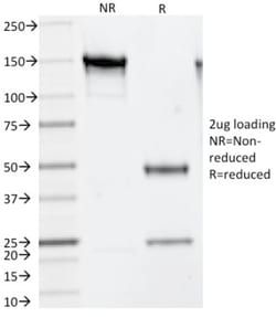
53 kDa
Clone
SPM565
Applications
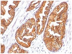
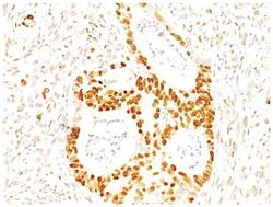
Western Blot, Flow Cytometry, Immunocytochemistry, Immunofluorescence, Immunohistochemistry (Paraffin), Immunohistochemistry (Frozen)
Conjugate
Unconjugated
Host Species
Mouse
Research Discipline
Cell Cycle and Replication
Formulation
PBS with 0.05% BSA. with 0.05% Sodium Azide
Gene Alias
ODCEC 4.1.1.17, ornithine decarboxylase, ornithine decarboxylase 1
Gene Symbols
ODC1
Isotype
IgG1 κ
Purification Method
Protein A purified
Test Specificity
Recognizes a 53kDa protein, identified as the Ornithine Decarboxylase (ODC-1). ODC is the initial and rate-limiting enzyme in the biosynthetic pathway of polyamines and is involved in the conversion of ornithine to putrescine. The biological activity of ODC-1 is rapidly induced in response to virtually all agents known to promote cell proliferation including hormones, drugs, growth factors, mitogens, and tumor promoters. Reportedly, ODC mRNA levels are elevated in lung carcinomas as well as in colon adenomas and carcinomas. ODC activity in colorectal carcinomas is greater than those in adenomas and normal mucosa.
Related Products


Description
- Description Ornithine Decarboxylase Monoclonal specifically detects Ornithine Decarboxylase in Human, Rat samples
- It is validated for Western Blot, Flow Cytometry, Immunohistochemistry, Immunocytochemistry/Immunofluorescence, Immunohistochemistry-Paraffin.
Compare Similar Items
Show Difference
Antigen: Ornithine Decarboxylase
Dilution: Western Blot 0.5-1ug/ml, Flow Cytometry 0.5-1ug/million cells, Immunocytochemistry/Immunofluorescence 0.5-1ug/ml, Immunohistochemistry-Paraffin 0.5-1.0ug/ml, Immunohistochemistry-Frozen 0.5-1.0ug/ml
Classification: Monoclonal
Form: Purified
Regulatory Status: RUO
Target Species: Human, Rat
Gene Accession No.: P11926
Gene ID (Entrez): 4953
Immunogen: Recombinant human ODC-1 protein
Primary or Secondary: Primary
Content And Storage: Store at 4C.
Molecular Weight of Antigen: 53 kDa
Clone: SPM565
Applications: Western Blot, Flow Cytometry, Immunocytochemistry, Immunofluorescence, Immunohistochemistry (Paraffin), Immunohistochemistry (Frozen)
Conjugate: Unconjugated
Host Species: Mouse
Research Discipline: Cell Cycle and Replication
Formulation: PBS with 0.05% BSA. with 0.05% Sodium Azide
Gene Alias: ODCEC 4.1.1.17, ornithine decarboxylase, ornithine decarboxylase 1
Gene Symbols: ODC1
Isotype: IgG1 κ
Purification Method: Protein A purified
Test Specificity: Recognizes a 53kDa protein, identified as the Ornithine Decarboxylase (ODC-1). ODC is the initial and rate-limiting enzyme in the biosynthetic pathway of polyamines and is involved in the conversion of ornithine to putrescine. The biological activity of ODC-1 is rapidly induced in response to virtually all agents known to promote cell proliferation including hormones, drugs, growth factors, mitogens, and tumor promoters. Reportedly, ODC mRNA levels are elevated in lung carcinomas as well as in colon adenomas and carcinomas. ODC activity in colorectal carcinomas is greater than those in adenomas and normal mucosa.
Antigen: p27/Kip1
Dilution: Western Blot 0.5-1ug/ml, Flow Cytometry 0.5-1ug/million cells, Immunocytochemistry/Immunofluorescence 0.5-1ug/ml, Immunohistochemistry-Paraffin 0.5-1.0ug/ml
Classification: Monoclonal
Form: Purified
Regulatory Status: RUO
Target Species: Human, Mouse, Rat, Primate
Gene Accession No.: P46527
Gene ID (Entrez): 1027
Immunogen: Purified GST-p27 fusion protein of human origin
Primary or Secondary: Primary
Content And Storage: Store at 4C.
Molecular Weight of Antigen: __
Clone: SX53G8
Applications: Western Blot, Flow Cytometry, Immunocytochemistry, Immunofluorescence, Immunohistochemistry (Paraffin)
Conjugate: Unconjugated
Host Species: Mouse
Research Discipline: Breast Cancer, Cancer, Cell Cycle and Replication, DNA Repair, Phospho Specific, Prostate Cancer, Tumor Suppressors
Formulation: PBS with 0.05% BSA. with 0.05% Sodium Azide
Gene Alias: CDKN4, cyclin-dependent kinase inhibitor 1B, cyclin-dependent kinase inhibitor 1B (p27, Kip1), Cyclin-dependent kinase inhibitor p27, KIP1P27KIP1, MEN1B, MEN4, p27Kip1
Gene Symbols: CDKN1B
Isotype: IgG1 κ
Purification Method: Protein A purified
Test Specificity: This MAb recognizes a 27kDa protein, identified as the p27Kip1, a cell cycle regulatory mitotic inhibitor. It is highly specific and shows no cross-reaction with other related mitotic inhibitors. In Western blotting of cell lysates from 7 human breast cancer cell lines (ZR75-1, ZR75-30, MCF-7, MDAMB453, T47D, CAL51, 734B), the antibody labels a single band corresponding to p27Kip1. It functions as a negative regulator of G1 progression and has been proposed to function as a possible mediator of TGF- induced G1 arrest. p27Kip1 is a candidate tumor suppressor gene. Reportedly, low p27 expression has been associated with unfavorable prognosis in renal cell carcinoma, colon carcinoma, breast carcinomas, non-small-cell lung carcinoma, hepatocellular carcinoma, multiple myeloma, and lymph node metastases in papillary carcinoma of the thyroid, as well as a more aggressive phenotype in carcinoma of the cervix.

Antigen: p27/Kip1
Dilution: Western Blot 0.5-1ug/ml, Flow Cytometry 0.5-1ug/million cells, Immunocytochemistry/Immunofluorescence 0.5-1ug/ml, Immunohistochemistry-Paraffin 0.5-1.0ug/ml
Classification: Monoclonal
Form: Purified
Regulatory Status: RUO
Target Species: Human, Mouse, Rat, Primate
Gene Accession No.: P46527
Gene ID (Entrez): 1027
Immunogen: Purified GST-p27 fusion protein of human origin
Primary or Secondary: Primary
Content And Storage: Store at 4C.
Molecular Weight of Antigen: __
Clone: SPM348
Applications: Western Blot, Flow Cytometry, Immunocytochemistry, Immunofluorescence, Immunohistochemistry (Paraffin)
Conjugate: Unconjugated
Host Species: Mouse
Research Discipline: Breast Cancer, Cancer, Cell Cycle and Replication, DNA Repair, Phospho Specific, Prostate Cancer, Tumor Suppressors
Formulation: PBS with 0.05% BSA. with 0.05% Sodium Azide
Gene Alias: CDKN4, cyclin-dependent kinase inhibitor 1B, cyclin-dependent kinase inhibitor 1B (p27, Kip1), Cyclin-dependent kinase inhibitor p27, KIP1P27KIP1, MEN1B, MEN4, p27Kip1
Gene Symbols: CDKN1B
Isotype: IgG1 κ
Purification Method: Protein A purified
Test Specificity: This MAb recognizes a 27kDa protein, identified as the p27Kip1, a cell cycle regulatory mitotic inhibitor. It is highly specific and shows no cross-reaction with other related mitotic inhibitors. p27Kip1 functions as a negative regulator of G1 progression and has been proposed to function as a possible mediator of TGF- induced G1 arrest. p27Kip1 is a candidate tumor suppressor gene. This MAb is excellent for staining of formalin-fixed tissues.