p53 Mouse, Clone: SPM589, Novus Biologicals™
Mouse Monoclonal Antibody
Manufacturer: Fischer Scientific
The price for this product is unavailable. Please request a quote
Antigen
p53
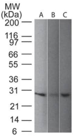
Dilution
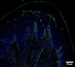
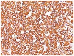
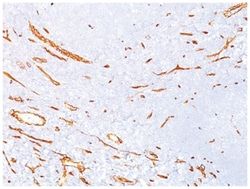
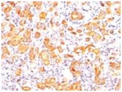
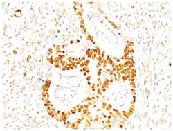
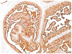
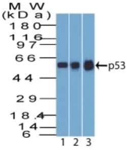
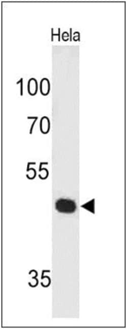
Western Blot 0.5-1ug/ml, Flow Cytometry 0.5-1ug/million cells, Immunocytochemistry/Immunofluorescence 1-2ug/ml, Immunohistochemistry-Paraffin 0.5-1.0ug/ml
Classification
Monoclonal
Form
Purified
Regulatory Status
RUO
Target Species
Human, Mouse (Negative), Rat (Negative)
Gene Accession No.
P04637
Gene ID (Entrez)
7157
Immunogen
Recombinant human wild-type p53 protein
Primary or Secondary
Primary
Content And Storage
Store at 4C.
Molecular Weight of Antigen
53 kDa
Clone
SPM589
Applications
Western Blot, Flow Cytometry, Immunocytochemistry, Immunofluorescence, Immunohistochemistry (Paraffin)
Conjugate
Unconjugated
Host Species
Mouse
Research Discipline
Apoptosis, Cancer, Cell Cycle and Replication, Cellular Markers, Checkpoint signaling, Core ESC Like Genes, DNA Double Strand Break Repair, DNA Repair, HIF Target Genes, Hypoxia, Neuroscience, Neurotransmission, p53 Pathway, Phospho Specific, Stem Cell Markers, Transcription Factors and Regulators, Tumor Suppressors
Formulation
PBS with 0.05% BSA. with 0.05% Sodium Azide
Gene Alias
Antigen NY-CO-13, FLJ92943, LFS1TRP53, p53, p53 tumor suppressor, P53cellular tumor antigen p53, Phosphoprotein p53, transformation-related protein 53, tumor protein p53, Tumor suppressor p53
Gene Symbols
TP53
Isotype
IgG2a
Purification Method
Protein A purified
Test Specificity
Recognizes a 53kDa protein, which is identified as p53 suppressor gene product. It reacts with the mutant as well as the wild form of p53 under denaturing and non-denaturing conditions. Its epitope maps within the N-terminus (aa 20-25) of p53 oncoprotein. p53 is a tumor suppressor gene expressed in a wide variety of tissue types and is involved in regulating cell growth, replication, and apoptosis. It binds to MDM2, SV40 T antigen and human papilloma virus E6 protein. Positive nuclear staining with p53 antibody has been reported to be a negative prognostic factor in breast carcinoma, lung carcinoma, colorectal, and urothelial carcinoma. Anti-p53 positivity has also been used to differentiate uterine serous carcinoma from endometrioid carcinoma as well as to detect intratubular germ cell neoplasia. Mutations involving p53 are found in a wide variety of malignant tumors, including breast, ovarian, bladder, colon, lung, and melanoma.
Related Products


Description
- Description p53 Monoclonal specifically detects p53 in Human, Mouse (Negative), Rat (Negative) samples
- It is validated for Western Blot, Flow Cytometry, Immunohistochemistry, Immunocytochemistry/Immunofluorescence, Immunohistochemistry-Paraffin.
Compare Similar Items
Show Difference
Antigen: p53
Dilution: Western Blot 0.5-1ug/ml, Flow Cytometry 0.5-1ug/million cells, Immunocytochemistry/Immunofluorescence 1-2ug/ml, Immunohistochemistry-Paraffin 0.5-1.0ug/ml
Classification: Monoclonal
Form: Purified
Regulatory Status: RUO
Target Species: Human, Mouse (Negative), Rat (Negative)
Gene Accession No.: P04637
Gene ID (Entrez): 7157
Immunogen: Recombinant human wild-type p53 protein
Primary or Secondary: Primary
Content And Storage: Store at 4C.
Molecular Weight of Antigen: 53 kDa
Clone: SPM589
Applications: Western Blot, Flow Cytometry, Immunocytochemistry, Immunofluorescence, Immunohistochemistry (Paraffin)
Conjugate: Unconjugated
Host Species: Mouse
Research Discipline: Apoptosis, Cancer, Cell Cycle and Replication, Cellular Markers, Checkpoint signaling, Core ESC Like Genes, DNA Double Strand Break Repair, DNA Repair, HIF Target Genes, Hypoxia, Neuroscience, Neurotransmission, p53 Pathway, Phospho Specific, Stem Cell Markers, Transcription Factors and Regulators, Tumor Suppressors
Formulation: PBS with 0.05% BSA. with 0.05% Sodium Azide
Gene Alias: Antigen NY-CO-13, FLJ92943, LFS1TRP53, p53, p53 tumor suppressor, P53cellular tumor antigen p53, Phosphoprotein p53, transformation-related protein 53, tumor protein p53, Tumor suppressor p53
Gene Symbols: TP53
Isotype: IgG2a
Purification Method: Protein A purified
Test Specificity: Recognizes a 53kDa protein, which is identified as p53 suppressor gene product. It reacts with the mutant as well as the wild form of p53 under denaturing and non-denaturing conditions. Its epitope maps within the N-terminus (aa 20-25) of p53 oncoprotein. p53 is a tumor suppressor gene expressed in a wide variety of tissue types and is involved in regulating cell growth, replication, and apoptosis. It binds to MDM2, SV40 T antigen and human papilloma virus E6 protein. Positive nuclear staining with p53 antibody has been reported to be a negative prognostic factor in breast carcinoma, lung carcinoma, colorectal, and urothelial carcinoma. Anti-p53 positivity has also been used to differentiate uterine serous carcinoma from endometrioid carcinoma as well as to detect intratubular germ cell neoplasia. Mutations involving p53 are found in a wide variety of malignant tumors, including breast, ovarian, bladder, colon, lung, and melanoma.
Antigen: Pax7
Dilution: Western Blot 0.5-1ug/ml, Flow Cytometry 0.5-1ug/million cells, Immunocytochemistry/Immunofluorescence 0.5-1ug/ml
Classification: Monoclonal
Form: Purified
Regulatory Status: RUO
Target Species: Human, Mouse, Rat, Chicken, Zebrafish
Gene Accession No.: P23759
Gene ID (Entrez): 5081
Immunogen: Recombinant fragment (aa300-600) of human PAX7 protein
Primary or Secondary: Primary
Content And Storage: Store at 4C.
Molecular Weight of Antigen: 57 kDa
Clone: PAX7/497
Applications: Western Blot, Flow Cytometry, Immunocytochemistry, Immunofluorescence
Conjugate: Unconjugated
Host Species: Mouse
Research Discipline: Apoptosis, Mesenchymal Stem Cell Markers, Stem Cell Markers
Formulation: PBS with 0.05% BSA. with 0.05% Sodium Azide
Gene Alias: FLJ37460, HuP1, paired box 7, paired box gene 7, paired box homeotic gene 7, paired box protein Pax-7, paired domain gene 7, PAX7 transcriptional factor, PAX7B, RMS2
Gene Symbols: PAX7
Isotype: IgG1 κ
Purification Method: Protein A purified
Test Specificity: The Pax gene family of nuclear transcription factors is comprised of nine members that function during embryogenesis to regulate the temporal and position-dependent differentiation of cells. In addition, the family is involved in a variety of signal transduction pathways in the adult organism. Mutations in the Pax family of proteins have been linked to disease and cancer in humans. Pax-7 is a protein specifically expressed in cultured satellite cell-derived myoblasts. In situ hybridization reveals that Pax-7 is also expressed in satellite cells residing in adult muscle. A chromosomal aberration in the gene encoding Pax-7 causes rhabdomyosarcoma 2 (RMS2) (also called alveolar rhabdomyosarcoma).
Antigen: UCH-L1/PGP9.5
Dilution: Western Blot 0.5-1ug/ml, Immunohistochemistry-Paraffin 0.5-1.0ug/ml
Classification: Monoclonal
Form: Purified
Regulatory Status: RUO
Target Species: Human, Mouse, Rat, Porcine, Bovine
Gene Accession No.: P09936
Gene ID (Entrez): 7345
Immunogen: Native UchL1 (PGP9.5) protein from brain
Primary or Secondary: Primary
Content And Storage: Store at 4C.
Molecular Weight of Antigen: __
Clone: SPM574
Applications: Western Blot, Immunohistochemistry (Paraffin)
Conjugate: Unconjugated
Host Species: Mouse
Research Discipline: Alzheimers Research, Cellular Markers, Neurodegeneration, Neuronal Cell Markers, Neuroscience, Neurotransmission
Formulation: PBS with 0.05% BSA. with 0.05% Sodium Azide
Gene Alias: EC 3.4.19.12, EC 6.-, Neuron cytoplasmic protein 9.5, PARK5, PGP 9.5, PGP9.5, PGP9.5Uch-L1, PGP95, ubiquitin carboxyl-terminal esterase L1 (ubiquitin thiolesterase), ubiquitin carboxyl-terminal hydrolase isozyme L1, ubiquitin C-terminal hydrolase, Ubiquitin thioesterase L1, UCHL1, UCH-L1
Gene Symbols: UCHL1
Isotype: IgG1 κ
Purification Method: Protein A purified
Test Specificity: This MAb reacts with a protein of 20-30kDa, identified as PGP9.5, also known as ubiquitin carboxyl-terminal hydrolase-1 (UchL1). Initially, PGP9.5 expression in normal tissues was reported in neurons and neuroendocrine cells but later it was found in distal renal tubular epithelium, spermatogonia, Leydig cells, oocytes, melanocytes, prostatic secretory epithelium, ejaculatory duct cells, epididymis, mammary epithelial cells, Merkel cells, and dermal fibroblasts. Furthermore, immunostaining for PGP9.5 has been shown in a wide variety of mesenchymal neoplasms as well. A mutation in PGP9.5 gene is believed to cause a form of Parkinson's disease.