7105414

Pax7 Mouse, Clone: PAX7/497, Novus Biologicals™

Mouse Monoclonal Antibody has been used in 4 publications

Manufacturer: Fischer Scientific

The price for this product is unavailable. Please request a quote

Antigen

Pax7

Dilution

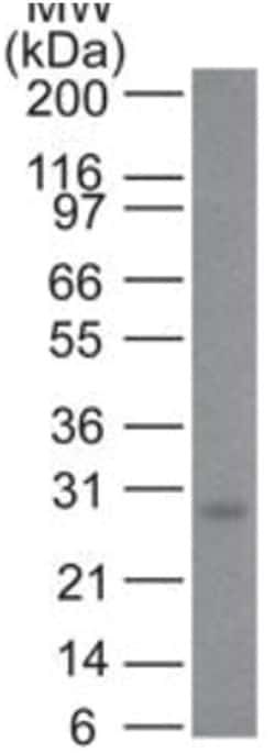
Western Blot 0.5-1ug/ml, Flow Cytometry 0.5-1ug/million cells, Immunocytochemistry/Immunofluorescence 0.5-1ug/ml

Classification

Monoclonal

Form

Purified

Regulatory Status

RUO

Target Species

Human, Mouse, Rat, Chicken, Zebrafish

Gene Accession No.

P23759

Gene ID (Entrez)

5081

Immunogen

Recombinant fragment (aa300-600) of human PAX7 protein

Primary or Secondary

Primary

Content And Storage

Store at 4C.

Molecular Weight of Antigen

57 kDa

Clone

PAX7/497

Applications

Western Blot, Flow Cytometry, Immunocytochemistry, Immunofluorescence

Conjugate

Unconjugated

Host Species

Mouse

Research Discipline

Apoptosis, Mesenchymal Stem Cell Markers, Stem Cell Markers

Formulation

PBS with 0.05% BSA. with 0.05% Sodium Azide

Gene Alias

FLJ37460, HuP1, paired box 7, paired box gene 7, paired box homeotic gene 7, paired box protein Pax-7, paired domain gene 7, PAX7 transcriptional factor, PAX7B, RMS2

Gene Symbols

PAX7

Isotype

IgG1 κ

Purification Method

Protein A purified

Test Specificity

The Pax gene family of nuclear transcription factors is comprised of nine members that function during embryogenesis to regulate the temporal and position-dependent differentiation of cells. In addition, the family is involved in a variety of signal transduction pathways in the adult organism. Mutations in the Pax family of proteins have been linked to disease and cancer in humans. Pax-7 is a protein specifically expressed in cultured satellite cell-derived myoblasts. In situ hybridization reveals that Pax-7 is also expressed in satellite cells residing in adult muscle. A chromosomal aberration in the gene encoding Pax-7 causes rhabdomyosarcoma 2 (RMS2) (also called alveolar rhabdomyosarcoma).

Related Products

Img

Fischer Scientific

7105479

Mouse Monoclonal Antibody...

Img

Fischer Scientific

7105399

Mouse Monoclonal Antibody...

Img

Fischer Scientific

7105622

Mouse Monoclonal Antibody...

Img

Fischer Scientific

7105622

Mouse Monoclonal Antibody...

Img

Fischer Scientific

7105405

Mouse Monoclonal Antibody has been ...

Img

Fischer Scientific

7105404

Mouse Monoclonal Antibody has been ...

Img

Fischer Scientific

7105619

Mouse Monoclonal Antibody...

Img

Fischer Scientific

7105490

Mouse Monoclonal Antibody...

Description

  • Pax7 Monoclonal specifically detects Pax7 in Human, Mouse, Rat, Chicken, Zebrafish samples
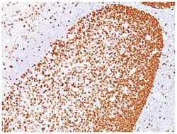
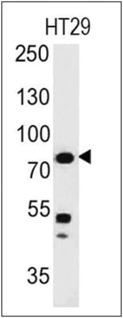
  • It is validated for Western Blot, Flow Cytometry, Immunocytochemistry/Immunofluorescence, Immunohistochemistry-Frozen.
Compare Similar Items

Show Difference

Img

Fischer Scientific

7105414

Mouse Monoclonal Antibody has been ...


Antigen:
Pax7

Dilution:
Western Blot 0.5-1ug/ml, Flow Cytometry 0.5-1ug/million cells, Immunocytochemistry/Immunofluorescence 0.5-1ug/ml

Classification:
Monoclonal

Form:
Purified

Regulatory Status:
RUO

Target Species:
Human, Mouse, Rat, Chicken, Zebrafish

Gene Accession No.:
P23759

Gene ID (Entrez):
5081

Immunogen:
Recombinant fragment (aa300-600) of human PAX7 protein

Primary or Secondary:
Primary

Content And Storage:
Store at 4C.

Molecular Weight of Antigen:
57 kDa

Clone:
PAX7/497

Applications:
Western Blot, Flow Cytometry, Immunocytochemistry, Immunofluorescence

Conjugate:
Unconjugated

Host Species:
Mouse

Research Discipline:
Apoptosis, Mesenchymal Stem Cell Markers, Stem Cell Markers

Formulation:
PBS with 0.05% BSA. with 0.05% Sodium Azide

Gene Alias:
FLJ37460, HuP1, paired box 7, paired box gene 7, paired box homeotic gene 7, paired box protein Pax-7, paired domain gene 7, PAX7 transcriptional factor, PAX7B, RMS2

Gene Symbols:
PAX7

Isotype:
IgG1 κ

Purification Method:
Protein A purified

Test Specificity:
The Pax gene family of nuclear transcription factors is comprised of nine members that function during embryogenesis to regulate the temporal and position-dependent differentiation of cells. In addition, the family is involved in a variety of signal transduction pathways in the adult organism. Mutations in the Pax family of proteins have been linked to disease and cancer in humans. Pax-7 is a protein specifically expressed in cultured satellite cell-derived myoblasts. In situ hybridization reveals that Pax-7 is also expressed in satellite cells residing in adult muscle. A chromosomal aberration in the gene encoding Pax-7 causes rhabdomyosarcoma 2 (RMS2) (also called alveolar rhabdomyosarcoma).

Img

Fischer Scientific

7105415

Mouse Monoclonal Antibody...


Antigen:
UCH-L1/PGP9.5

Dilution:
Western Blot 0.5-1ug/ml, Immunohistochemistry-Paraffin 0.5-1.0ug/ml

Classification:
Monoclonal

Form:
Purified

Regulatory Status:
RUO

Target Species:
Human, Mouse, Rat, Porcine, Bovine

Gene Accession No.:
P09936

Gene ID (Entrez):
7345

Immunogen:
Native UchL1 (PGP9.5) protein from brain

Primary or Secondary:
Primary

Content And Storage:
Store at 4C.

Molecular Weight of Antigen:
__

Clone:
SPM574

Applications:
Western Blot, Immunohistochemistry (Paraffin)

Conjugate:
Unconjugated

Host Species:
Mouse

Research Discipline:
Alzheimers Research, Cellular Markers, Neurodegeneration, Neuronal Cell Markers, Neuroscience, Neurotransmission

Formulation:
PBS with 0.05% BSA. with 0.05% Sodium Azide

Gene Alias:
EC 3.4.19.12, EC 6.-, Neuron cytoplasmic protein 9.5, PARK5, PGP 9.5, PGP9.5, PGP9.5Uch-L1, PGP95, ubiquitin carboxyl-terminal esterase L1 (ubiquitin thiolesterase), ubiquitin carboxyl-terminal hydrolase isozyme L1, ubiquitin C-terminal hydrolase, Ubiquitin thioesterase L1, UCHL1, UCH-L1

Gene Symbols:
UCHL1

Isotype:
IgG1 κ

Purification Method:
Protein A purified

Test Specificity:
This MAb reacts with a protein of 20-30kDa, identified as PGP9.5, also known as ubiquitin carboxyl-terminal hydrolase-1 (UchL1). Initially, PGP9.5 expression in normal tissues was reported in neurons and neuroendocrine cells but later it was found in distal renal tubular epithelium, spermatogonia, Leydig cells, oocytes, melanocytes, prostatic secretory epithelium, ejaculatory duct cells, epididymis, mammary epithelial cells, Merkel cells, and dermal fibroblasts. Furthermore, immunostaining for PGP9.5 has been shown in a wide variety of mesenchymal neoplasms as well. A mutation in PGP9.5 gene is believed to cause a form of Parkinson's disease.

Img

Fischer Scientific

7105416

Mouse Monoclonal Antibody...


Antigen:
UCH-L1/PGP9.5

Dilution:
Western Blot 0.5-1ug/ml, Immunocytochemistry/Immunofluorescence 1-2ug/ml, Immunocytochemistry 1:500

Classification:
Monoclonal

Form:
Purified

Regulatory Status:
RUO

Target Species:
Human, Mouse, Rat, Porcine, Bovine, Canine, Guinea Pig, Rabbit, Sheep, Zebrafish

Gene Accession No.:
P09936

Gene ID (Entrez):
7345

Immunogen:
Native UchL1 (PGP9.5) protein from brain

Primary or Secondary:
Primary

Content And Storage:
Store at 4C.

Molecular Weight of Antigen:
__

Clone:
SPM575

Applications:
Western Blot, Immunocytochemistry, Immunofluorescence

Conjugate:
Unconjugated

Host Species:
Mouse

Research Discipline:
Alzheimers Research, Cellular Markers, Neurodegeneration, Neuronal Cell Markers, Neuroscience, Neurotransmission

Formulation:
PBS with 0.05% BSA. with 0.05% Sodium Azide

Gene Alias:
EC 3.4.19.12, EC 6.-, Neuron cytoplasmic protein 9.5, PARK5, PGP 9.5, PGP9.5, PGP9.5Uch-L1, PGP95, ubiquitin carboxyl-terminal esterase L1 (ubiquitin thiolesterase), ubiquitin carboxyl-terminal hydrolase isozyme L1, ubiquitin C-terminal hydrolase, Ubiquitin thioesterase L1, UCHL1, UCH-L1

Gene Symbols:
UCHL1

Isotype:
IgG2a κ

Purification Method:
Protein A purified

Test Specificity:
This MAb reacts with a protein of 20-30kDa, identified as PGP9.5, also known as ubiquitin carboxyl-terminal hydrolase-1 (UchL1). Initially, PGP9.5 expression in normal tissues was reported in neurons and neuroendocrine cells but later it was found in distal renal tubular epithelium, spermatogonia, Leydig cells, oocytes, melanocytes, prostatic secretory epithelium, ejaculatory duct cells, epididymis, mammary epithelial cells, Merkel cells, and dermal fibroblasts. Furthermore, immunostaining for PGP9.5 has been shown in a wide variety of mesenchymal neoplasms as well. A mutation in PGP9.5 gene is believed to cause a form of Parkinson's disease.

Img

Fischer Scientific

7105417

Mouse Monoclonal Antibody...


Antigen:
PLGF

Dilution:
Western Blot 1-2ug/ml, SDS-Page

Classification:
Monoclonal

Form:
Purified

Regulatory Status:
RUO

Target Species:
Human

Gene Accession No.:
P49763

Gene ID (Entrez):
5228

Immunogen:
Recombinant human PLGF protein

Primary or Secondary:
Primary

Content And Storage:
Store at 4C.

Molecular Weight of Antigen:
18 kDa

Clone:
PLGF/94

Applications:
Western Blot, SDS-Page

Conjugate:
Unconjugated

Host Species:
Mouse

Research Discipline:
Angiogenesis

Formulation:
PBS with 0.05% BSA. with 0.05% Sodium Azide

Gene Alias:
D12S1900, PGFL, placenta growth factor, placental growth factor, placental growth factor, vascular endothelial growth factor-related protein, PlGF, PlGF-2, PLGFplacental growth factor-like, SHGC-10760

Gene Symbols:
PGF

Isotype:
IgG1 κ

Purification Method:
Protein A purified

Test Specificity:
The onset of angiogenesis is believed to be an early event in tumorigenesis and may facilitate tumor progression and metastasis. Several growth factors with angiogenic activity have been described. These include Fibroblast Growth Factor (FGF), Platelet Derived Growth Factor (PDGF), Vascular Endothelial Growth Factor (VEGF) and Placenta Growth Factor (PLGF). Placenta growth factor (PLGF) is a secreted protein primarily produced by placental trophoblasts but also expressed in other endothelial cells and tumors. There are three isoforms, PLGF-1, PLGF-2, and PLGF-3. PLGF-2 is expressed up until week 8 in the placenta; the placental tissues continuously express PLGF-1 and PLGF-3 but only PLGF-1 is found in colon and mammary carcinomas. PLGF acts to stimulate angiogenesis, endothelial growth and migration. PLGF is a powerful promoter of tumor growth and is upregulated in some cancers, and PLGF is thought to aid in atherosclerotic lesions and neovascular growth surrounding the lesion. Also, PLGF appears to aid aldosterone mediated atherosclerosis. Serum levels of PLGF in some cases are used as a potential biomarker for disease or genetic defect. Recent research indicates that PLGF levels are lower in mothers with Down syndrome fetuses. Evidence has suggested VEGF to be an obligatory component in PLGF signaling. While VEGF homodimers and VEGF/PLGF heterodimers function as potent mediators of mitogenic and chemotactic responses in endothelial cells, PLGF homodimers are effectual only at extremely high concentrations. Indeed, many of the physiological effects attributed to VEGF may actually be a result of VEGF/PLGF. VEGF and PLGF share a common receptor, Flt-1, and may also activate Flk-1/KDR.