PLGF Antibody (PLGF/94), Novus Biologicals™
Mouse Monoclonal Antibody
Manufacturer: Fischer Scientific
The price for this product is unavailable. Please request a quote
Antigen
PLGF
Dilution
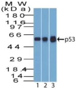
Western Blot 1-2ug/ml, SDS-Page
Classification
Monoclonal
Form
Purified
Regulatory Status
RUO
Target Species
Human
Gene Accession No.
P49763
Gene ID (Entrez)
5228
Immunogen
Recombinant human PLGF protein
Primary or Secondary
Primary
Content And Storage
Store at 4C.
Molecular Weight of Antigen
18 kDa
Clone
PLGF/94
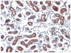
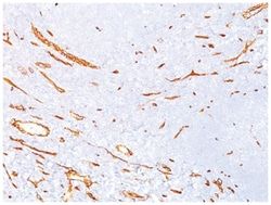
Applications
Western Blot, SDS-Page
Conjugate
Unconjugated
Host Species
Mouse
Research Discipline
Angiogenesis
Formulation
PBS with 0.05% BSA. with 0.05% Sodium Azide
Gene Alias
D12S1900, PGFL, placenta growth factor, placental growth factor, placental growth factor, vascular endothelial growth factor-related protein, PlGF, PlGF-2, PLGFplacental growth factor-like, SHGC-10760
Gene Symbols
PGF
Isotype
IgG1 κ
Purification Method
Protein A purified
Test Specificity
The onset of angiogenesis is believed to be an early event in tumorigenesis and may facilitate tumor progression and metastasis. Several growth factors with angiogenic activity have been described. These include Fibroblast Growth Factor (FGF), Platelet Derived Growth Factor (PDGF), Vascular Endothelial Growth Factor (VEGF) and Placenta Growth Factor (PLGF). Placenta growth factor (PLGF) is a secreted protein primarily produced by placental trophoblasts but also expressed in other endothelial cells and tumors. There are three isoforms, PLGF-1, PLGF-2, and PLGF-3. PLGF-2 is expressed up until week 8 in the placenta; the placental tissues continuously express PLGF-1 and PLGF-3 but only PLGF-1 is found in colon and mammary carcinomas. PLGF acts to stimulate angiogenesis, endothelial growth and migration. PLGF is a powerful promoter of tumor growth and is upregulated in some cancers, and PLGF is thought to aid in atherosclerotic lesions and neovascular growth surrounding the lesion. Also, PLGF appears to aid aldosterone mediated atherosclerosis. Serum levels of PLGF in some cases are used as a potential biomarker for disease or genetic defect. Recent research indicates that PLGF levels are lower in mothers with Down syndrome fetuses. Evidence has suggested VEGF to be an obligatory component in PLGF signaling. While VEGF homodimers and VEGF/PLGF heterodimers function as potent mediators of mitogenic and chemotactic responses in endothelial cells, PLGF homodimers are effectual only at extremely high concentrations. Indeed, many of the physiological effects attributed to VEGF may actually be a result of VEGF/PLGF. VEGF and PLGF share a common receptor, Flt-1, and may also activate Flk-1/KDR.
Related Products

Description
- PLGF Monoclonal specifically detects PLGF in Human samples
- It is validated for ELISA, Functional.
Compare Similar Items
Show Difference
Antigen: PLGF
Dilution: Western Blot 1-2ug/ml, SDS-Page
Classification: Monoclonal
Form: Purified
Regulatory Status: RUO
Target Species: Human
Gene Accession No.: P49763
Gene ID (Entrez): 5228
Immunogen: Recombinant human PLGF protein
Primary or Secondary: Primary
Content And Storage: Store at 4C.
Molecular Weight of Antigen: 18 kDa
Clone: PLGF/94
Applications: Western Blot, SDS-Page
Conjugate: Unconjugated
Host Species: Mouse
Research Discipline: Angiogenesis
Formulation: PBS with 0.05% BSA. with 0.05% Sodium Azide
Gene Alias: D12S1900, PGFL, placenta growth factor, placental growth factor, placental growth factor, vascular endothelial growth factor-related protein, PlGF, PlGF-2, PLGFplacental growth factor-like, SHGC-10760
Gene Symbols: PGF
Isotype: IgG1 κ
Purification Method: Protein A purified
Test Specificity: The onset of angiogenesis is believed to be an early event in tumorigenesis and may facilitate tumor progression and metastasis. Several growth factors with angiogenic activity have been described. These include Fibroblast Growth Factor (FGF), Platelet Derived Growth Factor (PDGF), Vascular Endothelial Growth Factor (VEGF) and Placenta Growth Factor (PLGF). Placenta growth factor (PLGF) is a secreted protein primarily produced by placental trophoblasts but also expressed in other endothelial cells and tumors. There are three isoforms, PLGF-1, PLGF-2, and PLGF-3. PLGF-2 is expressed up until week 8 in the placenta; the placental tissues continuously express PLGF-1 and PLGF-3 but only PLGF-1 is found in colon and mammary carcinomas. PLGF acts to stimulate angiogenesis, endothelial growth and migration. PLGF is a powerful promoter of tumor growth and is upregulated in some cancers, and PLGF is thought to aid in atherosclerotic lesions and neovascular growth surrounding the lesion. Also, PLGF appears to aid aldosterone mediated atherosclerosis. Serum levels of PLGF in some cases are used as a potential biomarker for disease or genetic defect. Recent research indicates that PLGF levels are lower in mothers with Down syndrome fetuses. Evidence has suggested VEGF to be an obligatory component in PLGF signaling. While VEGF homodimers and VEGF/PLGF heterodimers function as potent mediators of mitogenic and chemotactic responses in endothelial cells, PLGF homodimers are effectual only at extremely high concentrations. Indeed, many of the physiological effects attributed to VEGF may actually be a result of VEGF/PLGF. VEGF and PLGF share a common receptor, Flt-1, and may also activate Flk-1/KDR.
Antigen: Prolactin R
Dilution: Western Blot 0.25-0.5ug/ml, Flow Cytometry 0.5-1ug/million cells, Immunocytochemistry/Immunofluorescence 0.5-1ug/ml, Immunohistochemistry-Paraffin 0.5-1.0ug/ml
Classification: Monoclonal
Form: Purified
Regulatory Status: RUO
Target Species: Human
Gene Accession No.: P16471
Gene ID (Entrez): 5618
Immunogen: Semi-purified human prolactin receptor
Primary or Secondary: Primary
Content And Storage: Store at 4C.
Molecular Weight of Antigen: 70 kDa
Clone: SPM213
Applications: Western Blot, Flow Cytometry, Immunocytochemistry, Immunofluorescence, Immunohistochemistry (Paraffin)
Conjugate: Unconjugated
Host Species: Mouse
Research Discipline: Apoptosis
Formulation: PBS with 0.05% BSA. with 0.05% Sodium Azide
Gene Alias: delta 4-delta 7/11 truncated prolactin receptor, delta 4-SF1b truncated prolactin receptor, hPRL receptor, hPRLrI, PRL-R, prolactin receptor, prolactin receptor delta 7/11, secreted prolactin binding protein
Gene Symbols: PRLR
Isotype: IgG1 κ
Purification Method: Protein A purified
Test Specificity: It recognizes a protein of 70kDa, identified as prolactin receptor. Prolactin is a pituitary hormone involved in the stimulation of milk production, salt and water regulation, growth, development and reproduction. The initial step in its action is the binding to a specific membrane receptor (prolactin receptor), which belongs to the superfamily of class 1 cytokine receptors. The function of the prolactin receptor is mediated, at least in part, by two families of signaling molecules: Janus kinases and signal transducers and activators of transcription.