Cytokeratin 19 Antibody (A53-B/A2.26 + BA17), Novus Biologicals™
Mouse Monoclonal Antibody
Manufacturer: Fischer Scientific
The price for this product is unavailable. Please request a quote
Antigen
Cytokeratin 19
Dilution
Western Blot 0.5-1ug/ml, Flow Cytometry 0.5-1ug/million cells, Immunocytochemistry/Immunofluorescence 1-2ug/ml, Immunoprecipitation 1-2ug/500ug protein, Immunohistochemistry-Paraffin 0.5-1ug/ml, Immunohistochemistry-Frozen 0.5-1ug/ml
Classification
Monoclonal
Form
Purified
Regulatory Status
RUO
Target Species
Human
Gene Accession No.
P08727
Gene ID (Entrez)
3880
Immunogen
Human breast cancer MCF-7 cells (A53-B/A2); Human mammary epithelial organoids (BA17)
Primary or Secondary
Primary
Content And Storage
Store at 4C.
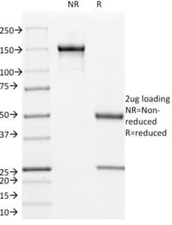
Molecular Weight of Antigen
40 kDa
Clone
A53-B/A2.26 + BA17
Applications
Western Blot, Flow Cytometry, Immunocytochemistry, Immunofluorescence, Immunoprecipitation, Immunohistochemistry (Paraffin)
Conjugate
Unconjugated
Host Species
Mouse
Research Discipline
Cancer, Cell Biology, Cellular Markers, Cytoskeleton Markers, Neuroscience, Stem Cell Markers
Formulation
PBS with 0.05% BSA. with 0.05% Sodium Azide
Gene Alias
CK19, CK-19, cytokeratin 19, Cytokeratin-19,40-kDa keratin intermediate filament, K19cytokeratin-19, K1CS, keratin 19, keratin, type I cytoskeletal 19, keratin, type I, 40-kd, keratin-19, MGC15366
Gene Symbols
KRT19
Isotype
IgG2a κ, IgG1 κ
Purification Method
Protein A purified
Test Specificity
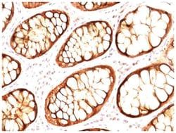
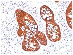
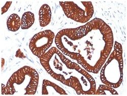
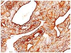
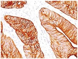
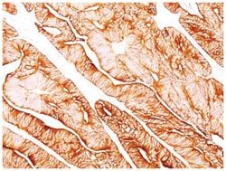
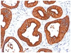
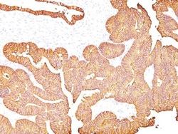
This MAb reacts with the rod domain of human cytokeratin-19 (CK19), a polypeptide of 40kDa. CK19 is expressed in sweat gland, mammary gland ductal and secretory cells, bile ducts, gastrointestinal tract, bladder urothelium, oral epithelia, esophagus, and ectocervical epithelium. Anti-CK19 reacts with a wide variety of epithelial malignancies including adenocarcinomas of the colon, stomach, pancreas, biliary tract, liver, and breast. Perhaps the most useful application is the identification of thyroid carcinoma of the papillary type, although 50%-60% of follicular carcinomas are also labeled. Anti-CK19 is a useful marker for detection of tumor cells in lymph nodes, peripheral blood, bone marrow and breast cancer.
Related Products


Description
- Cytokeratin 19 Monoclonal specifically detects Cytokeratin 19 in Human samples
- It is validated for Western Blot, Immunohistochemistry, Immunohistochemistry-Paraffin.
Compare Similar Items
Show Difference
Antigen: Cytokeratin 19
Dilution: Western Blot 0.5-1ug/ml, Flow Cytometry 0.5-1ug/million cells, Immunocytochemistry/Immunofluorescence 1-2ug/ml, Immunoprecipitation 1-2ug/500ug protein, Immunohistochemistry-Paraffin 0.5-1ug/ml, Immunohistochemistry-Frozen 0.5-1ug/ml
Classification: Monoclonal
Form: Purified
Regulatory Status: RUO
Target Species: Human
Gene Accession No.: P08727
Gene ID (Entrez): 3880
Immunogen: Human breast cancer MCF-7 cells (A53-B/A2); Human mammary epithelial organoids (BA17)
Primary or Secondary: Primary
Content And Storage: Store at 4C.
Molecular Weight of Antigen: 40 kDa
Clone: A53-B/A2.26 + BA17
Applications: Western Blot, Flow Cytometry, Immunocytochemistry, Immunofluorescence, Immunoprecipitation, Immunohistochemistry (Paraffin)
Conjugate: Unconjugated
Host Species: Mouse
Research Discipline: Cancer, Cell Biology, Cellular Markers, Cytoskeleton Markers, Neuroscience, Stem Cell Markers
Formulation: PBS with 0.05% BSA. with 0.05% Sodium Azide
Gene Alias: CK19, CK-19, cytokeratin 19, Cytokeratin-19,40-kDa keratin intermediate filament, K19cytokeratin-19, K1CS, keratin 19, keratin, type I cytoskeletal 19, keratin, type I, 40-kd, keratin-19, MGC15366
Gene Symbols: KRT19
Isotype: IgG2a κ, IgG1 κ
Purification Method: Protein A purified
Test Specificity: This MAb reacts with the rod domain of human cytokeratin-19 (CK19), a polypeptide of 40kDa. CK19 is expressed in sweat gland, mammary gland ductal and secretory cells, bile ducts, gastrointestinal tract, bladder urothelium, oral epithelia, esophagus, and ectocervical epithelium. Anti-CK19 reacts with a wide variety of epithelial malignancies including adenocarcinomas of the colon, stomach, pancreas, biliary tract, liver, and breast. Perhaps the most useful application is the identification of thyroid carcinoma of the papillary type, although 50%-60% of follicular carcinomas are also labeled. Anti-CK19 is a useful marker for detection of tumor cells in lymph nodes, peripheral blood, bone marrow and breast cancer.
Antigen: L1CAM
Dilution: Western Blot 0.5-1.0ug/ml, Flow Cytometry 0.5-1ug/million cells, Immunocytochemistry/Immunofluorescence 1-2ug/ml, Immunoprecipitation 1-2ug/500ug protein, Immunohistochemistry-Paraffin, Immunohistochemistry-Frozen 1-2ug/ml, SDS-Page
Classification: Monoclonal
Form: Purified
Regulatory Status: RUO
Target Species: Human
Gene Accession No.: P32004
Gene ID (Entrez): 3897
Immunogen: Homogenous suspension of 16 week human fetal brain
Primary or Secondary: Primary
Content And Storage: Store at 4C.
Molecular Weight of Antigen: 230 kDa
Clone: UJ127
Applications: Western Blot, Flow Cytometry, Immunocytochemistry, Immunofluorescence, Immunoprecipitation, Immunohistochemistry (Paraffin)
Conjugate: Unconjugated
Host Species: Mouse
Research Discipline: Cellular Markers, Glia Markers, Neuroscience
Formulation: PBS with 0.05% BSA. with 0.05% Sodium Azide
Gene Alias: antigen identified by monoclonal R1, CAML1N-CAML1, CD171, CD171 antigen, HSAS, HSAS1, L1 cell adhesion molecule, MASA, MIC5, N-CAM-L1, NCAM-L1, neural cell adhesion molecule L1, S10, SPG1
Gene Symbols: L1CAM
Isotype: IgG1 κ
Purification Method: Protein A purified
Test Specificity: Recognizes a cell surface protein of 220-240kDa, identified as L1 cell adhesion molecule. The L1CAM gene, which is located in Xq28, is involved in three distinct conditions: 1) HSAS (hydrocephalus-stenosis of the aqueduct of Sylvius); 2) MASA (mental retardation, aphasia, shuffling gait, and adducted thumbs); and 3) SPG1 (spastic paraplegia). The L1, neural cell adhesion molecule (L1CAM) also plays an important role in axon growth, fasciculation, and neural migration as well as in mediating neuronal differentiation. Expression of L1 protein is restricted to tissues arising from neuroectoderm. This MAb is useful in the identification of primitive neuroectodermal tumors. It binds to tumors of neuroectodermal and glial origin e.g. neuroblastoma and Schwannomas. It does not bind to pediatric or adult brain.
Antigen: EpCAM/TROP1
Dilution: Western Blot 0.25-0.5ug/ml, Flow Cytometry 0.5-1ug/million cells, Immunocytochemistry/Immunofluorescence 1-2ug/ml, Immunoprecipitation 1-2ug/500ug protein, Immunohistochemistry-Paraffin 0.5-1ug/ml, Immunohistochemistry-Frozen 0.5-1ug/ml
Classification: Monoclonal
Form: Purified
Regulatory Status: RUO
Target Species: Human, Rat (Negative)
Gene Accession No.: P16422
Gene ID (Entrez): 4072
Immunogen: MCF-7 human breast cancer cells
Primary or Secondary: Primary
Content And Storage: Store at 4C.
Molecular Weight of Antigen: 42 kDa
Clone: 323/A3
Applications: Western Blot, Flow Cytometry, Immunocytochemistry, Immunofluorescence, Immunoprecipitation, Immunohistochemistry (Paraffin)
Conjugate: Unconjugated
Host Species: Mouse
Research Discipline: Cancer
Formulation: PBS with 0.05% BSA. with 0.05% Sodium Azide
Gene Alias: 17-1A, 323/A3, ACSTD1, antigen identified by monoclonal AUA1, CD326 antigen, Cell surface glycoprotein Trop-1, chromosome 4, surface marker (35kD glycoprotein), DIAR5, EGP, EGP-2, EGP314, EGP40, EpCAM, epithelial cell adhesion molecule, Epithelial cell surface antigen, Epithelial glycoprotein, Epithelial glycoprotein 314, ESA, GA733-2EGP34, hEGP314, HNPCC8, KS 1/4 antigen, KS1/4, KSAHEA125, M1S2, M4S1Ly74, Major gastrointestinal tumor-associated protein GA733-2, MIC18MH99, MOC31, TACST-1, TACSTD1, TROP1CD326, Tumor-associated calcium signal transducer 1CO-17A
Gene Symbols: EPCAM
Isotype: IgG1 κ
Purification Method: Protein A purified
Test Specificity: EGP40 is a 40-43kDa transmembrane epithelial glycoprotein, also identified as epithelial specific antigen (ESA), or epithelial cellular adhesion molecule (Ep-CAM). It is expressed on baso-lateral cell surface in most simple epithelia and a vast majority of carcinomas. This antibody has been used to distinguish adenocarcinoma from pleural mesothelioma and hepatocellular carcinoma. This antibody is also useful in distinguishing serous carcinomas of the ovary from mesothelioma.