7105647

MLH1 Mouse anti-Human, Clone: MLH1/1324, Novus Biologicals™

Mouse Monoclonal Antibody

Manufacturer: Fischer Scientific

The price for this product is unavailable. Please request a quote

Antigen

MLH1

Dilution

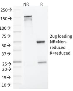
Western Blot 0.5-1.0ug/ml, SDS-Page

Classification

Monoclonal

Form

Purified

Regulatory Status

RUO

Target Species

Human

Gene Alias

COCA2FCC2, DNA mismatch repair protein Mlh1, hMLH1, HNPCC, HNPCC2MGC5172, mutL (E. coli) homolog 1 (colon cancer, nonpolyposis type 2), mutL homolog 1, colon cancer, nonpolyposis type 2 (E. coli), MutL protein homolog 1

Gene Symbols

MLH1

Isotype

IgG2b

Purification Method

Protein A or G purified

Test Specificity

This MAb recognizes a protein of 83kDa, identified as MLH1. Its epitope maps in aa 380-410. Defects in MLH1 are the cause of hereditary non-polyposis colorectal cancer type 2 (HNPCC2). Heterodimerizes with PMS2 to form MutL alpha, a component of the post-replicative DNA mismatch repair system (MMR). DNA repair is initiated by MutS alpha (MSH2-MSH6) or MutS beta (MSH2-MSH6) binding to a dsDNA mismatch, then MutL alpha is recruited to the heteroduplex. Assembly of the MutL-MutS-heteroduplex ternary complex in presence of RFC and PCNA is sufficient to activate endonuclease activity of PMS2. It introduces single-strand breaks near the mismatch and thus generates new entry points for the exonuclease EXO1 to degrade the strand containing the mismatch. DNA methylation would prevent cleavage and therefore assure that only the newly mutated DNA strand is going to be corrected. MutL alpha (MLH1-PMS2) interacts physically with the clamp loader subunits of DNA polymerase III, suggesting that it ma

Clone

MLH1/1324

Applications

Western Blot, SDS-Page

Conjugate

Unconjugated

Host Species

Mouse

Research Discipline

Apoptosis, Breast Cancer, Cancer, Cell Cycle and Replication, Checkpoint signaling, DNA Double Strand Break Repair, DNA Repair, Mismatch Repair, Tumor Suppressors

Formulation

10mM PBS and 0.05% BSA with 0.05% Sodium Azide

Gene ID (Entrez)

4292

Immunogen

Recombinant human MLH1 protein

Primary or Secondary

Primary

Content And Storage

Store at 4C.

Molecular Weight of Antigen

85 kDa

Related Products

Img

Fischer Scientific

7105674

Mouse Monoclonal Antibody...

Img

Fischer Scientific

7105675

Mouse Monoclonal Antibody...

Img

Fischer Scientific

7105717

Mouse Monoclonal Antibody...

Img

Fischer Scientific

7105411

Mouse Monoclonal Antibody...

Img

Fischer Scientific

7105842

Mouse Monoclonal Antibody...

Img

Fischer Scientific

7105842

Mouse Monoclonal Antibody...

Img

Fischer Scientific

7105841

Mouse Monoclonal Antibody...

Img

Fischer Scientific

7105655

Mouse Monoclonal Antibody...

Description

  • MLH1 Monoclonal specifically detects MLH1 in Human samples
  • It is validated for ELISA.

Compare Similar Items

Show Difference

Img

Fischer Scientific

7105647

Mouse Monoclonal Antibody...


Antigen:
MLH1

Dilution:
Western Blot 0.5-1.0ug/ml, SDS-Page

Classification:
Monoclonal

Form:
Purified

Regulatory Status:
RUO

Target Species:
Human

Gene Alias:
COCA2FCC2, DNA mismatch repair protein Mlh1, hMLH1, HNPCC, HNPCC2MGC5172, mutL (E. coli) homolog 1 (colon cancer, nonpolyposis type 2), mutL homolog 1, colon cancer, nonpolyposis type 2 (E. coli), MutL protein homolog 1

Gene Symbols:
MLH1

Isotype:
IgG2b

Purification Method:
Protein A or G purified

Test Specificity:
This MAb recognizes a protein of 83kDa, identified as MLH1. Its epitope maps in aa 380-410. Defects in MLH1 are the cause of hereditary non-polyposis colorectal cancer type 2 (HNPCC2). Heterodimerizes with PMS2 to form MutL alpha, a component of the post-replicative DNA mismatch repair system (MMR). DNA repair is initiated by MutS alpha (MSH2-MSH6) or MutS beta (MSH2-MSH6) binding to a dsDNA mismatch, then MutL alpha is recruited to the heteroduplex. Assembly of the MutL-MutS-heteroduplex ternary complex in presence of RFC and PCNA is sufficient to activate endonuclease activity of PMS2. It introduces single-strand breaks near the mismatch and thus generates new entry points for the exonuclease EXO1 to degrade the strand containing the mismatch. DNA methylation would prevent cleavage and therefore assure that only the newly mutated DNA strand is going to be corrected. MutL alpha (MLH1-PMS2) interacts physically with the clamp loader subunits of DNA polymerase III, suggesting that it ma

Clone:
MLH1/1324

Applications:
Western Blot, SDS-Page

Conjugate:
Unconjugated

Host Species:
Mouse

Research Discipline:
Apoptosis, Breast Cancer, Cancer, Cell Cycle and Replication, Checkpoint signaling, DNA Double Strand Break Repair, DNA Repair, Mismatch Repair, Tumor Suppressors

Formulation:
10mM PBS and 0.05% BSA with 0.05% Sodium Azide

Gene ID (Entrez):
4292

Immunogen:
Recombinant human MLH1 protein

Primary or Secondary:
Primary

Content And Storage:
Store at 4C.

Molecular Weight of Antigen:
85 kDa

Img

Fischer Scientific

7105651

Mouse Monoclonal Antibody...


Antigen:
pan Actin

Dilution:
Western Blot : 0.5 - 1.0 ug/ml, Flow Cytometry : 0.5 - 1 ug/million cells in 0.1 ml, Immunohistochemistry-Paraffin : 0.25 - 0.5 ug/ml, Immunofluorescence : 0.5 - 1.0 ug/ml, CyTOF-ready

Classification:
Monoclonal

Form:
Purified

Regulatory Status:
RUO

Target Species:
Human, Rat, Rabbit

Gene Alias:
ACTA, actin, alpha 1, skeletal muscle, alpha skeletal muscle, alpha skeletal muscle actin, alpha-actin-1, ASMA, CFTD, CFTDM, MPFD, NEM1, NEM2, NEM3

Gene Symbols:
ACTA1

Isotype:
IgG1 κ

Purification Method:
Protein A or G purified

Test Specificity:
This antibody recognizes actin of skeletal, cardiac, and smooth muscle cells. It is not reactive with other mesenchymal cells except for myoepithelium. Actin can be resolved on the basis of its isoelectric points into three distinctive components: alpha, beta, and gamma in order of increasing isoelectric point. Anti-muscle specific actin recognizes alpha and gamma isotypes of all muscle groups. Non-muscle cells such as vascular endothelial cells and connective tissues are non-reactive. Also, neoplastic cells of non-muscle-derived tissue such as carcinomas, melanomas, and lymphomas are negative.AIt stains tumors of smooth muscle (leiomyomas and leiomyosarcomas) as well as skeletal muscle (rhabdomyomas and rhabdomyosarcomas).

Clone:
MSA/953

Applications:
Western Blot, Flow Cytometry, Immunohistochemistry (Paraffin), Immunofluorescence, CyTOF

Conjugate:
Unconjugated

Host Species:
Mouse

Research Discipline:
Angiogenesis, Cancer, Cell Biology, Cellular Markers, Cytoskeleton Markers, Stem Cell Markers

Formulation:
PBS with No Preservative

Gene ID (Entrez):
58

Immunogen:
Synthetic peptide common among human muscle actins

Primary or Secondary:
Primary

Content And Storage:
Store at 4C short term. Aliquot and store at -20C long term. Avoid freeze-thaw cycles.

Molecular Weight of Antigen:
__

Img

Fischer Scientific

7105653

Mouse Monoclonal Antibody...


Antigen:
NF-H

Dilution:
Flow Cytometry : 0.5 - 1 ug/million cells in 0.1 ml, Immunohistochemistry-Paraffin : 0.25 - 0.5 ug/ml, CyTOF-ready

Classification:
Monoclonal

Form:
Purified

Regulatory Status:
RUO

Target Species:
Human, Mouse, Rat, Porcine, Chicken

Gene Alias:
200 kDa neurofilament protein, KIAA0845, Neurofilament Heavy (200kDa), neurofilament heavy polypeptide, Neurofilament triplet H protein, neurofilament, heavy polypeptide, neurofilament, heavy polypeptide 200kDa, NF200, NFH, NF-H

Gene Symbols:
NEFH

Isotype:
IgG

Purification Method:
Protein A or G purified

Test Specificity:
This MAb reacts with a 200kDa and 68kDa protein, identified as heavy and light sub-units of neurofilaments (NF-H & NF-L). Neurofilaments make up the main structural elements of axons and dendrites and are found in neurons, peripheral nerves, and sympathetic ganglion cells. Neurofilaments consist of three major subunits with molecular weights of 68kDa (NF-L), 160kDa (NF-M) and 200kDa (NF-H). Anti-neurofilament stains a number of neural, neuroendocrine, and endocrine tumors. Neuromas, ganglioneuromas, gangliogliomas, ganglioneuroblastomas, and neuroblastomas stain positively for anti-neurofilament. Neurofilaments are also present in paragangliomas as well as adrenal and extra-adrenal pheochromocytomas. Carcinoids, neuroendocrine carcinomas of the skin, and oat cell carcinomas of the lung also express neurofilament.

Clone:
RT-97 + NR-4

Applications:
Flow Cytometry, Immunohistochemistry (Paraffin), CyTOF

Conjugate:
Unconjugated

Host Species:
Mouse

Research Discipline:
Autophagy, Cellular Markers, Cytoskeleton Markers, Neurodegeneration, Neurofilaments, Neuronal Cell Markers, Neuroscience

Formulation:
PBS with No Preservative

Gene ID (Entrez):
4744

Immunogen:
Triton-X 100 insoluble proteins of rat brain (RT-97); Neurofilaments from porcine spinal cord (NR-4)

Primary or Secondary:
Primary

Content And Storage:
Store at 4C short term. Aliquot and store at -20C long term. Avoid freeze-thaw cycles.

Molecular Weight of Antigen:
__