IgG Antibody (B33/20) - Azide and BSA Free, Novus Biologicals™
Mouse Monoclonal Antibody
Manufacturer: Fischer Scientific
The price for this product is unavailable. Please request a quote
Antigen
IgG
Concentration
1.0 mg/mL
Applications
Flow Cytometry, Immunohistochemistry (Paraffin), Immunofluorescence, CyTOF
Conjugate
Unconjugated
Host Species
Mouse
Research Discipline
Immunology
Formulation
PBS with No Preservative
Gene ID (Entrez)
3500
Immunogen
Purified polyclonal human Ig Gamma Chain
Primary or Secondary
Primary
Content And Storage
Store at 4C short term. Aliquot and store at -20C long term. Avoid freeze-thaw cycles.
Molecular Weight of Antigen
75 kDa
Clone
B33/20
Dilution
Flow Cytometry : 0.5 - 1 ug/million cells in 0.1 ml, Immunohistochemistry-Paraffin : 0.5 - 1.0 ug/ml, Immunofluorescence : 0.5 - 1.0 ug/ml, CyTOF-ready
Classification
Monoclonal
Form
Purified
Regulatory Status
RUO
Target Species
Human
Gene Alias
immunoglobulin heavy constant gamma 1 (G1m marker)
Gene Symbols
IGHG
Isotype
IgG1 κ
Purification Method
Protein A or G purified
Test Specificity
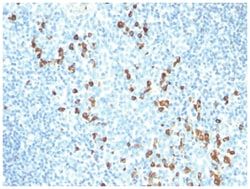
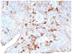
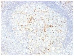
Recognizes a protein of 75kDa, identified as gamma heavy chain of human immunoglobulins. Its epitope maps in CH2 domain of Fc region of IgG. It reacts with all sub-classes of gamma chain of human immunoglobulins. It does not cross-react with alpha (IgA), u (IgM), epsilon (IgE), or delta (IgD), heavy chains, T-cells, monocytes, granulocytes, or erythrocytes. This MAb is useful in the identification of leukemias, plasmacytomas, and certain non-Hodgkin's lymphomas. The most common feature of these malignancies is the restricted expression of a single heavy chain class. Demonstration of clonality in lymphoid infiltrates indicates that the infiltrate is clonal and therefore malignant.
Related Products

Description
- IgG Monoclonal specifically detects IgG in Human samples
- It is validated for Flow Cytometry, Immunohistochemistry, Immunocytochemistry/Immunofluorescence, Immunohistochemistry-Paraffin, Immunofluorescence, CyTOF-ready.
Compare Similar Items
Show Difference
Antigen: IgG
Concentration: 1.0 mg/mL
Applications: Flow Cytometry, Immunohistochemistry (Paraffin), Immunofluorescence, CyTOF
Conjugate: Unconjugated
Host Species: Mouse
Research Discipline: Immunology
Formulation: PBS with No Preservative
Gene ID (Entrez): 3500
Immunogen: Purified polyclonal human Ig Gamma Chain
Primary or Secondary: Primary
Content And Storage: Store at 4C short term. Aliquot and store at -20C long term. Avoid freeze-thaw cycles.
Molecular Weight of Antigen: 75 kDa
Clone: B33/20
Dilution: Flow Cytometry : 0.5 - 1 ug/million cells in 0.1 ml, Immunohistochemistry-Paraffin : 0.5 - 1.0 ug/ml, Immunofluorescence : 0.5 - 1.0 ug/ml, CyTOF-ready
Classification: Monoclonal
Form: Purified
Regulatory Status: RUO
Target Species: Human
Gene Alias: immunoglobulin heavy constant gamma 1 (G1m marker)
Gene Symbols: IGHG
Isotype: IgG1 κ
Purification Method: Protein A or G purified
Test Specificity: Recognizes a protein of 75kDa, identified as gamma heavy chain of human immunoglobulins. Its epitope maps in CH2 domain of Fc region of IgG. It reacts with all sub-classes of gamma chain of human immunoglobulins. It does not cross-react with alpha (IgA), u (IgM), epsilon (IgE), or delta (IgD), heavy chains, T-cells, monocytes, granulocytes, or erythrocytes. This MAb is useful in the identification of leukemias, plasmacytomas, and certain non-Hodgkin's lymphomas. The most common feature of these malignancies is the restricted expression of a single heavy chain class. Demonstration of clonality in lymphoid infiltrates indicates that the infiltrate is clonal and therefore malignant.
Antigen: CD25/IL-2 R alpha
Concentration: 1.0 mg/mL
Applications: Flow Cytometry, Immunofluorescence, CyTOF
Conjugate: Unconjugated
Host Species: Mouse
Research Discipline: Adaptive Immunity, Apoptosis, B Cell Development and Differentiation Markers, Cytokine Research, Diabetes Research, Immunology, Innate Immunity, Mast Cell Markers, Signal Transduction
Formulation: PBS with No Preservative
Gene ID (Entrez): 3559
Immunogen: Stimulated human leukocytes
Primary or Secondary: Primary
Content And Storage: Store at 4C short term. Aliquot and store at -20C long term. Avoid freeze-thaw cycles.
Molecular Weight of Antigen: 55 kDa
Clone: 143-13
Dilution: Flow Cytometry : 0.5 - 1 ug/million cells in 0.1 ml, Immunofluorescence : 0.5 - 1.0 ug/ml, CyTOF-ready
Classification: Monoclonal
Form: Purified
Regulatory Status: RUO
Target Species: Human
Gene Alias: CD25, CD25 antigen, IDDM10, IL-2 receptor subunit alpha, IL2R, IL-2R subunit alpha, IL-2-RA, IL2-RA, interleukin 2 receptor, alpha, interleukin-2 receptor subunit alpha, p55, TAC antigen, TCGFR
Gene Symbols: IL2RA
Isotype: IgG1 κ
Purification Method: Protein A or G purified
Test Specificity: Recognizes a protein of 55kDa, identified as CD25. It is expressed on activated T- and B-cells and activated monocytes/macrophages. With respect to lymphomas, CD25 is present on malignant cells of Hodgkin's disease, HTLV-1+ adult T-cell leukemia, cutaneous T-cell lymphoma, and hair cell leukemia. Increased levels of soluble CD25 are observed in the leukemias/lymphomas and inflammatory/ autoimmune diseases. CD25 molecule alone appears to function as a low affinity receptor and associates with CD122 (IL-2R chain, p75) and CD132 (common chain) to form the high affinity IL-2 receptor complex. CD25 antibodies detect three epitope regions, A, B and C. This MAb recognizes the epitope B, which is located at residue 3-104 of CD25 and doe not block IL-2 binding to CD25.