PMEL17/SILV Antibody (PMEL/783) - Azide and BSA Free, Novus Biologicals™
Mouse Monoclonal Antibody
Manufacturer: Fischer Scientific
The price for this product is unavailable. Please request a quote
Antigen
PMEL17/SILV
Concentration
1.0 mg/mL
Applications
Flow Cytometry, Immunohistochemistry (Paraffin), Immunofluorescence, CyTOF
Conjugate
Unconjugated
Host Species
Mouse
Target Species
Human
Gene Alias
D12S53EP1, gp100, ME20, ME20-M, melanocyte protein mel 17, Melanocyte protein Pmel 17, Melanocytes lineage-specific antigen GP100, Melanoma-associated ME20 antigen, melanosomal matrix protein17, PMEL17P100, premelanosome proteinME20M, SI, SIL, silver (mouse homolog) like, silver homolog (mouse), Silver locus protein homolog, silver, mouse, homolog of, SILVPmel17
Gene Symbols
PMEL
Isotype
IgG1 κ
Purification Method
Protein A or G purified
Test Specificity
Cytotoxic T lymphocytes (CTLs) recognize melanoma-associated antigens, which belong to three main groups. These groups include tumor-associated testis-specific antigens, melanocyte differentiation antigens and mutated or aberrantly expressed antigens, which are routinely used as markers to identify melanomas based on their binding to specific monoclonal antibodies. gp100, also designated ME20-M, ME20-S and PMEL 17, is classified as a melanocyte differentiation antigen and is expressed at low levels in normal cell lines and tissues, but is upregulated in melanocytes. gp100 is a highly glycosylated protein. It is also the product of proteolytic cleavage, which results in a secreted protein
Clone
PMEL/783
Dilution
Flow Cytometry : 0.5 - 1 ug/million cells in 0.1 ml, Immunohistochemistry-Paraffin : 0.5 - 1.0 ug/ml, Immunofluorescence : 0.5 - 1.0 ug/ml, CyTOF-ready
Classification
Monoclonal
Form
Purified
Regulatory Status
RUO
Formulation
PBS with No Preservative
Gene ID (Entrez)
6490
Immunogen
Recombinant human SILV protein
Primary or Secondary
Primary
Content And Storage
Store at 4C short term. Aliquot and store at -20C long term. Avoid freeze-thaw cycles.
Molecular Weight of Antigen
95 kDa
Related Products

Description
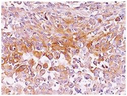
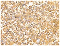
- PMEL17/SILV Monoclonal specifically detects PMEL17/SILV in Human samples
- It is validated for Western Blot, Immunohistochemistry, Immunohistochemistry-Paraffin.
Compare Similar Items
Show Difference
Antigen: PMEL17/SILV
Concentration: 1.0 mg/mL
Applications: Flow Cytometry, Immunohistochemistry (Paraffin), Immunofluorescence, CyTOF
Conjugate: Unconjugated
Host Species: Mouse
Target Species: Human
Gene Alias: D12S53EP1, gp100, ME20, ME20-M, melanocyte protein mel 17, Melanocyte protein Pmel 17, Melanocytes lineage-specific antigen GP100, Melanoma-associated ME20 antigen, melanosomal matrix protein17, PMEL17P100, premelanosome proteinME20M, SI, SIL, silver (mouse homolog) like, silver homolog (mouse), Silver locus protein homolog, silver, mouse, homolog of, SILVPmel17
Gene Symbols: PMEL
Isotype: IgG1 κ
Purification Method: Protein A or G purified
Test Specificity: Cytotoxic T lymphocytes (CTLs) recognize melanoma-associated antigens, which belong to three main groups. These groups include tumor-associated testis-specific antigens, melanocyte differentiation antigens and mutated or aberrantly expressed antigens, which are routinely used as markers to identify melanomas based on their binding to specific monoclonal antibodies. gp100, also designated ME20-M, ME20-S and PMEL 17, is classified as a melanocyte differentiation antigen and is expressed at low levels in normal cell lines and tissues, but is upregulated in melanocytes. gp100 is a highly glycosylated protein. It is also the product of proteolytic cleavage, which results in a secreted protein
Clone: PMEL/783
Dilution: Flow Cytometry : 0.5 - 1 ug/million cells in 0.1 ml, Immunohistochemistry-Paraffin : 0.5 - 1.0 ug/ml, Immunofluorescence : 0.5 - 1.0 ug/ml, CyTOF-ready
Classification: Monoclonal
Form: Purified
Regulatory Status: RUO
Formulation: PBS with No Preservative
Gene ID (Entrez): 6490
Immunogen: Recombinant human SILV protein
Primary or Secondary: Primary
Content And Storage: Store at 4C short term. Aliquot and store at -20C long term. Avoid freeze-thaw cycles.
Molecular Weight of Antigen: 95 kDa
Antigen: PMEL17/SILV
Concentration: 1.0 mg/mL
Applications: Flow Cytometry, Immunohistochemistry (Paraffin), Immunofluorescence, CyTOF
Conjugate: Unconjugated
Host Species: Mouse
Target Species: Human
Gene Alias: D12S53EP1, gp100, ME20, ME20-M, melanocyte protein mel 17, Melanocyte protein Pmel 17, Melanocytes lineage-specific antigen GP100, Melanoma-associated ME20 antigen, melanosomal matrix protein17, PMEL17P100, premelanosome proteinME20M, SI, SIL, silver (mouse homolog) like, silver homolog (mouse), Silver locus protein homolog, silver, mouse, homolog of, SILVPmel17
Gene Symbols: PMEL
Isotype: IgG
Purification Method: Protein A or G purified
Test Specificity: By immunohistochemistry, it specifically recognizes a protein in melanocytes and melanomas. This MAb reacts with junctional and blue nevus cells and variably with fetal and neonatal melanocytes. Intradermal nevi, normal adult melanocytes, and non-melanocytic cells are negative. It does not stain tumor cells of epithelial, lymphoid, glial, or mesenchymal origin. Metastatic amelanotic melanoma can often be confused with a variety of poorly differentiated carcinomas, large cell lymphomas, and sarcomas using H & E stains alone. It is also difficult to differentiate melanoma from spindle cell carcinomas and various types of mesenchymal neoplasms. This MAb stains fetal and neonatal melanocytes, junctional and blue nevus cells, and malignant melanoma. This MAb also stains Angiomyolipoma (PEComa).
Clone: HMB45 + PMEL/783
Dilution: Flow Cytometry : 0.5 - 1 ug/million cells in 0.1 ml, Immunohistochemistry-Paraffin : 0.5 - 1.0 ug/ml, Immunofluorescence : 0.5 - 1.0 ug/ml, CyTOF-ready
Classification: Monoclonal
Form: Purified
Regulatory Status: RUO
Formulation: PBS with No Preservative
Gene ID (Entrez): 6490
Immunogen: Extract of pigmented melanoma metastases from lymph nodes (HMB45); Recombinant human SILV protein (PMEL/783)
Primary or Secondary: Primary
Content And Storage: Store at 4C short term. Aliquot and store at -20C long term. Avoid freeze-thaw cycles.
Molecular Weight of Antigen: 95 kDa
Antigen: p21/CIP1/CDKN1A
Concentration: 1.0 mg/mL
Applications: Western Blot, Flow Cytometry, Immunohistochemistry (Paraffin), Immunofluorescence, CyTOF
Conjugate: Unconjugated
Host Species: Mouse
Target Species: Human, Mouse (Negative), Rat (Negative)
Gene Alias: CAP20cyclin-dependent kinase inhibitor 1, CDK-interacting protein 1, CDKN1melanoma differentiation associated protein 6, CIP1WAF1CDK-interaction protein 1, cyclin-dependent kinase inhibitor 1A (p21, Cip1), MDA6, MDA-6, Melanoma differentiation-associated protein 6, p21, p21CIP1, p21Cip1/Waf1, PIC1, SDI1DNA synthesis inhibitor, wild-type p53-activated fragment 1
Gene Symbols: CDKN1A
Isotype: IgG2a κ
Purification Method: Protein A or G purified
Test Specificity: This MAb recognizes a 21kDa protein, identified as the p21WAF1 tumor suppressor protein. This MAb is highly specific to p21 and shows no cross-reaction with other closely related mitotic inhibitors. p21WAF1 is a specific inhibitor of cdk s and a tumor suppressor involved in the pathogenesis of a variety of malignancies. The expression of this gene acts as an inhibitor of the cell cycle during G1 phase and is tightly controlled by the tumor suppressor protein p53. Its expression is induced by the wild type, but not mutant, p53 suppressor protein. Normal cells generally display a rather intense nuclear p21 expression. Loss of p21 expression has been reported in many carcinomas (gastric carcinoma, non-small cell lung carcinoma, thyroid carcinoma).
Clone: SPM306
Dilution: Western Blot : 1 - 2 ug/ml, Flow Cytometry : 0.5 - 1 ug/million cells in 0.1 ml, Immunohistochemistry-Paraffin : 2 - 4 ug/ml, Immunofluorescence : 1 - 2 ug/ml, CyTOF-ready
Classification: Monoclonal
Form: Purified
Regulatory Status: RUO
Formulation: PBS with No Preservative
Gene ID (Entrez): 1026
Immunogen: Human recombinant p21 protein
Primary or Secondary: Primary
Content And Storage: Store at 4C short term. Aliquot and store at -20C long term. Avoid freeze-thaw cycles.
Molecular Weight of Antigen: 21 kDa