CD31/PECAM-1 Antibody (C31.3 + C31.7 + C31.10) - Azide and BSA Free, Novus Biologicals™
Mouse Monoclonal Antibody has been used in 4 publications
Manufacturer: Fischer Scientific
The price for this product is unavailable. Please request a quote
Antigen
CD31/PECAM-1
Concentration
1.0 mg/mL
Applications
Western Blot, Flow Cytometry, Immunohistochemistry (Paraffin), Immunofluorescence, CyTOF
Conjugate
Unconjugated
Host Species
Mouse
Research Discipline
Angiogenesis, Cancer, Cellular Markers, Cytoskeleton Markers, Embryonic Stem Cell Markers, Endothelial Cell Markers, Extracellular Matrix, Hematopoietic Stem Cell Markers, Immunology, Mesenchymal Stem Cell Markers, Myeloid Cell Markers, Myeloid derived Suppressor Cell, Signal Transduction, Stem Cell Markers
Formulation
PBS with No Preservative
Gene ID (Entrez)
5175
Immunogen
Human recombinant CD31 protein
Primary or Secondary
Primary
Content And Storage
Store at 4C short term. Aliquot and store at -20C long term. Avoid freeze-thaw cycles.
Clone
C31.3 + C31.7 + C31.10
Dilution
Western Blot : 0.5 - 1.0 ug/ml, Flow Cytometry : 0.5 - 1 ug/million cells in 0.1 ml, Immunohistochemistry-Paraffin : 1 - 2 ug/ml, Immunofluorescence : 0.5 - 1.0 ug/ml, CyTOF-ready
Classification
Monoclonal
Form
Purified
Regulatory Status
RUO
Target Species
Human, Rat, Cynomolgus Monkey, Rabbit
Gene Alias
adhesion molecule, CD31, CD31 antigen, CD31/EndoCAM, EndoCAM, FLJ34100, FLJ58394, GPIIA', PECA1, PECAM-1, PECAM-1, CD31/EndoCAM, platelet endothelial cell adhesion molecule, platelet/endothelial cell adhesion molecule
Gene Symbols
PECAM1
Isotype
IgG
Purification Method
Protein A or G purified
Test Specificity
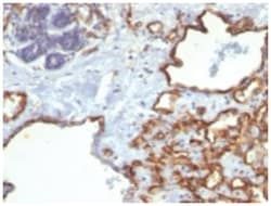
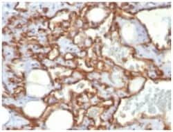
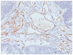
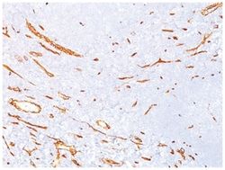
CD31 (PECAM-1) is a transmembrane glycoprotein member of the immunoglobulin supergene family of adhesion molecules. CD31 is expressed by stem cells of the hematopoietic system and is primarily used to identify and concentrate these cells for experimental studies as well as for bone marrow transplantation. Anti-CD31 has shown to be highly specific and sensitive for vascular endothelial cells. Staining of nonvascular tumors (excluding hematopoietic neoplasms) is rare. CD31 MAb reacts with normal, benign, and malignant endothelial cells which make up blood vessel lining. The level of CD31 expression can help to determine the degree of tumor angiogenesis, and a high level of CD31 expression may imply a rapidly growing tumor and potentially a predictor of tumor recurrence.
Related Products

Description
- CD31/PECAM-1 Monoclonal specifically detects CD31/PECAM-1 in Human, Rat, Cynomolgus Monkey, Rabbit samples
- It is validated for Western Blot, Immunohistochemistry, Immunocytochemistry/Immunofluorescence, Immunohistochemistry-Paraffin.
Compare Similar Items
Show Difference
Antigen: CD31/PECAM-1
Concentration: 1.0 mg/mL
Applications: Western Blot, Flow Cytometry, Immunohistochemistry (Paraffin), Immunofluorescence, CyTOF
Conjugate: Unconjugated
Host Species: Mouse
Research Discipline: Angiogenesis, Cancer, Cellular Markers, Cytoskeleton Markers, Embryonic Stem Cell Markers, Endothelial Cell Markers, Extracellular Matrix, Hematopoietic Stem Cell Markers, Immunology, Mesenchymal Stem Cell Markers, Myeloid Cell Markers, Myeloid derived Suppressor Cell, Signal Transduction, Stem Cell Markers
Formulation: PBS with No Preservative
Gene ID (Entrez): 5175
Immunogen: Human recombinant CD31 protein
Primary or Secondary: Primary
Content And Storage: Store at 4C short term. Aliquot and store at -20C long term. Avoid freeze-thaw cycles.
Clone: C31.3 + C31.7 + C31.10
Dilution: Western Blot : 0.5 - 1.0 ug/ml, Flow Cytometry : 0.5 - 1 ug/million cells in 0.1 ml, Immunohistochemistry-Paraffin : 1 - 2 ug/ml, Immunofluorescence : 0.5 - 1.0 ug/ml, CyTOF-ready
Classification: Monoclonal
Form: Purified
Regulatory Status: RUO
Target Species: Human, Rat, Cynomolgus Monkey, Rabbit
Gene Alias: adhesion molecule, CD31, CD31 antigen, CD31/EndoCAM, EndoCAM, FLJ34100, FLJ58394, GPIIA', PECA1, PECAM-1, PECAM-1, CD31/EndoCAM, platelet endothelial cell adhesion molecule, platelet/endothelial cell adhesion molecule
Gene Symbols: PECAM1
Isotype: IgG
Purification Method: Protein A or G purified
Test Specificity: CD31 (PECAM-1) is a transmembrane glycoprotein member of the immunoglobulin supergene family of adhesion molecules. CD31 is expressed by stem cells of the hematopoietic system and is primarily used to identify and concentrate these cells for experimental studies as well as for bone marrow transplantation. Anti-CD31 has shown to be highly specific and sensitive for vascular endothelial cells. Staining of nonvascular tumors (excluding hematopoietic neoplasms) is rare. CD31 MAb reacts with normal, benign, and malignant endothelial cells which make up blood vessel lining. The level of CD31 expression can help to determine the degree of tumor angiogenesis, and a high level of CD31 expression may imply a rapidly growing tumor and potentially a predictor of tumor recurrence.
Antigen: EMI1
Concentration: 1.0 mg/mL
Applications: Western Blot, Flow Cytometry, Immunohistochemistry (Paraffin), Immunofluorescence, CyTOF
Conjugate: Unconjugated
Host Species: Mouse
Research Discipline: __
Formulation: PBS with No Preservative
Gene ID (Entrez): 26271
Immunogen: Recombinant fragment (203 amino acid residues between aa 1-250) of human EMI1 protein
Primary or Secondary: Primary
Content And Storage: Store at 4C short term. Aliquot and store at -20C long term. Avoid freeze-thaw cycles.
Clone: EMI1/1176
Dilution: Western Blot : 1 - 2 ug/ml, Flow Cytometry : 0.5 - 1 ug/million cells in 0.1 ml, Immunohistochemistry-Paraffin : 0.5 - 1.0 ug/ml, Immunofluorescence : 0.5 - 1.0 ug/ml, CyTOF-ready
Classification: Monoclonal
Form: Purified
Regulatory Status: RUO
Target Species: Human
Gene Alias: EMI1Early mitotic inhibitor 1, F-box only protein 5, F-box protein 5, FBX5F-box protein Fbx5, Fbxo31
Gene Symbols: FBXO5
Isotype: IgG2a κ
Purification Method: Protein A or G purified
Test Specificity: It recognizes a 56kDa protein, which is identified as Early Mitotic Inhibitor-1 (EMI1). It regulates mitosis by inhibiting the anaphase promoting complex/cyclosome (APC). Emi1 is a conserved F box protein containing a zinc-binding region essential for APC inhibition. The Emi1 protein functions to promote cyclin A accumulation and S phase entry in somatic cells by inhibiting the APC complex. At the G1-S transition, Emi1 is transcriptionally induced by the E2F transcription factor. Emi1 overexpression accelerates S-phase entry and can override a G1 block caused by overexpression of Cdh1 or the E2F-inhibitor p105 retinoblastoma protein (pRb). Depleting cells of Emi1 through RNA interference prevents accumulation of cyclin A and inhibits S phase entry.
Antigen: beta-1,3-Glucuronyltransferase 1/B3GAT1
Concentration: 1.0 mg/mL
Applications: Immunohistochemistry (Paraffin), Immunofluorescence
Conjugate: Unconjugated
Host Species: Mouse
Research Discipline: Cancer, Immunology
Formulation: PBS with No Preservative
Gene ID (Entrez): 27087
Immunogen: Human peripheral blood mononuclear cells
Primary or Secondary: Primary
Content And Storage: Store at 4C short term. Aliquot and store at -20C long term. Avoid freeze-thaw cycles.
Clone: SPM527
Dilution: Immunohistochemistry-Paraffin : 2 - 4 ug/ml, Immunofluorescence : 0.5 - 1.0 ug/ml
Classification: Monoclonal
Form: Purified
Regulatory Status: RUO
Target Species: Human, Rat (Negative)
Gene Alias: Beta-1,3-glucuronyltransferase 1, beta-1,3-glucuronyltransferase 1 (glucuronosyltransferase P), EC 2.4.1.135, galactosylgalactosylxylosylprotein 3-beta-glucuronosyltransferase 1, GlcAT-P, GLCATPGLCUATP, glcUAT-P, Glucuronosyltransferase P, NK1, NK-1, UDP-GlcUA:glycoprotein beta-1,3-glucuronyltransferase
Gene Symbols: B3GAT1
Isotype: IgM κ
Purification Method: Protein A or G purified
Test Specificity: Anti-CD57 marks a subset of lymphocytes known as natural killer (NK) cells. Follicular center cell lymphomas often contain many NK cells within the neoplastic follicles. Anti-CD57 also stains neuroendocrine cells and their derived tumors, including carcinoid tumor and medulloblastoma. Anti-CD57 can also be useful in separating type B3 thymoma from thymic carcinoma when combined with a panel that includes antibodies against GLUT1, CD5, and CEA.