Cytokeratin 5/8 Antibody (C-50) - Azide and BSA Free, Novus Biologicals™
Mouse Monoclonal Antibody
Manufacturer: Fischer Scientific
The price for this product is unavailable. Please request a quote
Antigen
Cytokeratin 5/8
Concentration
1.0 mg/mL
Applications
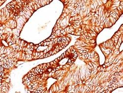
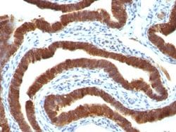
Western Blot, Flow Cytometry, Immunohistochemistry (Paraffin), Immunofluorescence, CyTOF
Conjugate
Unconjugated
Host Species
Mouse
Target Species
Human, Mouse, Rat, Porcine, Canine, Ferret, Hamster, Primate, Sheep, Chicken (Negative), Xenopus (Negative)
Gene Alias
cell proliferation-inducing protein 46, CK18, CK5, CYK18, DDD, K18, K1CO, K5, Keratin, Keratin 18, Keratin 5, KRT18, krt5, KRT5A, KRTB, type I cytoskeletal 18, type II cytoskeletal 5
Gene Symbols
KRT5
Isotype
IgG1 κ
Purification Method
Protein A or G purified
Test Specificity
It reacts with keratin 5 (58kDa) and keratin 8 (52.5kDa). Simple epithelia express cytokeratin 8 in combination with 18. On the other hand, basal cells of stratified epithelia express cytokeratin 5 paired with 14. This antibody therefore, reacts with a wide range of epithelia and their carcinomas.
Clone
C-50
Dilution
Western Blot : 0.5 - 1.0 ug/ml, Flow Cytometry : 0.5 - 1 ug/million cells in 0.1 ml, Immunohistochemistry-Paraffin : 0.5 - 1.0 ug/ml, Immunofluorescence : 0.5 - 1.0 ug/ml, CyTOF-ready
Classification
Monoclonal
Form
Purified
Regulatory Status
RUO
Formulation
PBS with No Preservative
Gene ID (Entrez)
3852
Immunogen
Cytoskeletal preparation from HeLa cells
Primary or Secondary
Primary
Content And Storage
Store at 4C short term. Aliquot and store at -20C long term. Avoid freeze-thaw cycles.
Related Products


Description
- Cytokeratin 5/8 Monoclonal specifically detects Cytokeratin 5/8 in Human, Mouse, Rat, Porcine, Bovine, Canine, Hamster, Ferret, Sheep, Chicken (Negative), Xenopus (Negative) samples
- It is validated for Western Blot, Flow Cytometry, Immunohistochemistry, Immunocytochemistry/Immunofluorescence, Immunohistochemistry-Paraffin, Immunofluorescence, CyTOF-ready.
Compare Similar Items
Show Difference
Antigen: Cytokeratin 5/8
Concentration: 1.0 mg/mL
Applications: Western Blot, Flow Cytometry, Immunohistochemistry (Paraffin), Immunofluorescence, CyTOF
Conjugate: Unconjugated
Host Species: Mouse
Target Species: Human, Mouse, Rat, Porcine, Canine, Ferret, Hamster, Primate, Sheep, Chicken (Negative), Xenopus (Negative)
Gene Alias: cell proliferation-inducing protein 46, CK18, CK5, CYK18, DDD, K18, K1CO, K5, Keratin, Keratin 18, Keratin 5, KRT18, krt5, KRT5A, KRTB, type I cytoskeletal 18, type II cytoskeletal 5
Gene Symbols: KRT5
Isotype: IgG1 κ
Purification Method: Protein A or G purified
Test Specificity: It reacts with keratin 5 (58kDa) and keratin 8 (52.5kDa). Simple epithelia express cytokeratin 8 in combination with 18. On the other hand, basal cells of stratified epithelia express cytokeratin 5 paired with 14. This antibody therefore, reacts with a wide range of epithelia and their carcinomas.
Clone: C-50
Dilution: Western Blot : 0.5 - 1.0 ug/ml, Flow Cytometry : 0.5 - 1 ug/million cells in 0.1 ml, Immunohistochemistry-Paraffin : 0.5 - 1.0 ug/ml, Immunofluorescence : 0.5 - 1.0 ug/ml, CyTOF-ready
Classification: Monoclonal
Form: Purified
Regulatory Status: RUO
Formulation: PBS with No Preservative
Gene ID (Entrez): 3852
Immunogen: Cytoskeletal preparation from HeLa cells
Primary or Secondary: Primary
Content And Storage: Store at 4C short term. Aliquot and store at -20C long term. Avoid freeze-thaw cycles.
Antigen: Cytokeratin 10
Concentration: 1.0 mg/mL
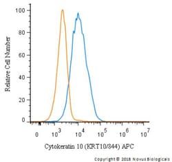
Applications: Flow Cytometry, Immunohistochemistry (Paraffin), Immunofluorescence, CyTOF
Conjugate: Unconjugated
Host Species: Mouse
Target Species: Human, Mouse
Gene Alias: BCIE, BIE, CK10, CK-10, cytokeratin 10, Cytokeratin-10, EHK, K10keratosis palmaris et plantaris, keratin 10, Keratin 10 (epidermolytic hyperkeratosis; keratosis palmaris et plantaris), keratin, type I cytoskeletal 10, keratin-10, KPP
Gene Symbols: KRT10
Isotype: IgG1 κ
Purification Method: Protein A or G purified
Test Specificity: This MAb recognizes a protein of 56.5kDa, identified as cytokeratin 10 (CK10). CK10 is expressed in all suprabasal layers of the epidermis. In the epidermis, expression of CK10 strictly parallels the extent of differentiation; it is absent in the basal layer, appears in the first suprabasal layers and increases in concentration towards the granular layer. However, CK10 is rarely detected in early stages of vulvar squamous carcinomas (tumors less than 2 cm, clinical stage I) regardless of the tumor grade. In larger and more advanced tumors (greater than 2 cm, clinical stages II and III), CK10 is detected very frequently. Expression of CK10 is related to maturation of malignant keratinocytes, being preferentially detected in more-differentiated parts.
Clone: KRT10/844
Dilution: Flow Cytometry : 0.5 - 1 ug/million cells in 0.1 ml, Immunohistochemistry-Paraffin : 0.1 - 0.2 ug/ml, Immunofluorescence : 0.5 - 1.0 ug/ml, CyTOF-ready
Classification: Monoclonal
Form: Purified
Regulatory Status: RUO
Formulation: PBS with No Preservative
Gene ID (Entrez): 3858
Immunogen: Recombinant human KRT10 protein
Primary or Secondary: Primary
Content And Storage: Store at 4C short term. Aliquot and store at -20C long term. Avoid freeze-thaw cycles.
Antigen: Fc gamma RIIA/CD32a
Concentration: 1.0 mg/mL
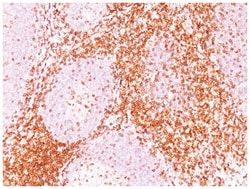
Applications: Flow Cytometry, Immunofluorescence, CyTOF
Conjugate: Unconjugated
Host Species: Mouse
Target Species: Human
Gene Alias: CD32 antigen, CD32A, CD32MGC23887, CDw32fc-gamma-RIIa, Fc fragment of IgG, low affinity IIa, receptor (CD32), Fc fragment of IgG, low affinity IIa, receptor for (CD32), FCG2, Fc-gamma RII-a, Fc-gamma-RIIa, FcGR, FCGR2, FCGR2A1, FcRII-a, IGFR2MGC30032, IgG Fc receptor II-a, Immunoglobulin G Fc receptor II, low affinity immunoglobulin gamma Fc region receptor II-a
Gene Symbols: FCGR2A
Isotype: IgG1 κ
Purification Method: Protein A or G purified
Test Specificity: This MAb reacts with a CD32 (FcgRII) epitope distinct from that defined by MAb 8.26 and the epitope overlaps with that of MAb 7.30 (cluster 4). It displays a stronger reaction with Daudi than with U937 cells. The epitope is located in domain 2 of FcgRIIa. Its Fab'2 fragments block immune complex binding. CD32 (Fc?RII) is a type 1 transmembrane glycoprotein that mediates several functions including phagocytosis, cytotoxicity, and immunomodulation as well as platelet aggregation. Three genes (A, B, and C) encode CD32 and at least 6 isoforms are generated via alternative mRNA splicing, i.e., IIa1, IIa2, IIb1, IIb2, IIb3 and IIc. Monocytes/macrophages, placental trophoblasts and endothelial cells express all isoforms. In addition, the IIb isoform is expressed by B cells, and the IIa isoform by platelets, granulocytes and, weakly, by B cells. NK cells and neutrophils express Isoform IIc. CD32 binds weakly to the Fc region of monomeric IgG but more strongly to IgG aggregates and immune complexes.
Clone: 8.7
Dilution: Flow Cytometry : 0.5 - 1 ug/million cells in 0.1 ml, Immunofluorescence : 0.5 - 1.0 ug/ml, CyTOF-ready
Classification: Monoclonal
Form: Purified
Regulatory Status: RUO
Formulation: PBS with No Preservative
Gene ID (Entrez): 2212
Immunogen: K562 and FcgRII+L cells
Primary or Secondary: Primary
Content And Storage: Store at 4C short term. Aliquot and store at -20C long term. Avoid freeze-thaw cycles.