Neprilysin/CD10 Antibody (CB-CALLA) - Azide and BSA Free, Novus Biologicals™
Mouse Monoclonal Antibody
Manufacturer: Fischer Scientific
The price for this product is unavailable. Please request a quote
Antigen
Neprilysin/CD10
Concentration
1.0 mg/mL
Applications
Flow Cytometry, Immunofluorescence, CyTOF
Conjugate
Unconjugated
Host Species
Mouse
Research Discipline
B Cell Development and Differentiation Markers, Cancer, Cell Biology, Cytokine Research, Immunology, Stem Cell Markers
Formulation
PBS with No Preservative
Gene ID (Entrez)
4311
Immunogen
Human CD10
Primary or Secondary
Primary
Content And Storage
Store at 4C short term. Aliquot and store at -20C long term. Avoid freeze-thaw cycles.
Molecular Weight of Antigen
100 kDa
Clone
CB-CALLA
Dilution
Flow Cytometry : 0.5 - 1 ug/million cells in 0.1 ml, Immunofluorescence : 0.5 - 1.0 ug/ml, CyTOF-ready
Classification
Monoclonal
Form
Purified
Regulatory Status
RUO
Target Species
Human
Gene Alias
Atriopeptidase, CALLAmembrane metallo-endopeptidase (neutral endopeptidase, enkephalinase), CD10 antigen, CD10), CD10membrane metallo-endopeptidase variant 1, Common acute lymphocytic leukemia antigen, DKFZp686O16152, EC 3.4.24, EC 3.4.24.11, Enkephalinase, EPN, membrane metallo-endopeptidase, membrane metallo-endopeptidase variant 2, MGC126681, MGC126707, NEPmembrane metallo-endopeptidase (neutral endopeptidase, enkephalinase, CALLA, neprilysin, Neutral endopeptidase, Neutral endopeptidase 24.11, SFE, Skin fibroblast elastase
Gene Symbols
MME
Isotype
IgG2b κ
Purification Method
Protein A or G purified
Test Specificity
Recognizes a 100kDa glycoprotein, identified as CD10, also known as Common Acute Lymphocytic Leukemia Antigen (CALLA). It is a cell surface enzyme with neutral metalloendopeptidase activity, which inactivates a variety of biologically active peptides. CD10 is expressed on the cells of lymphoblastic, Burkitt's, and follicular germinal center lymphomas, and on cells from patients with chronic myelocytic leukemia (CML). It is also expressed on the surface of normal early lymphoid progenitor cells, immature B cells within adult bone marrow and germinal center B cells within lymphoid tissue. CD10 is also present on breast myoepithelial cells, bile canaliculi, fibroblasts, with especially high expression on the brush border of kidney and gut epithelial cells.
Description
- Description Neprilysin/CD10 Monoclonal specifically detects Neprilysin/CD10 in Human samples
- It is validated for Flow Cytometry, Immunocytochemistry/Immunofluorescence, Immunofluorescence, CyTOF-ready.
Compare Similar Items
Show Difference

Antigen: Neprilysin/CD10
Concentration: 1.0 mg/mL
Applications: Flow Cytometry, Immunofluorescence, CyTOF
Conjugate: Unconjugated
Host Species: Mouse
Research Discipline: B Cell Development and Differentiation Markers, Cancer, Cell Biology, Cytokine Research, Immunology, Stem Cell Markers
Formulation: PBS with No Preservative
Gene ID (Entrez): 4311
Immunogen: Human CD10
Primary or Secondary: Primary
Content And Storage: Store at 4C short term. Aliquot and store at -20C long term. Avoid freeze-thaw cycles.
Molecular Weight of Antigen: 100 kDa
Clone: CB-CALLA
Dilution: Flow Cytometry : 0.5 - 1 ug/million cells in 0.1 ml, Immunofluorescence : 0.5 - 1.0 ug/ml, CyTOF-ready
Classification: Monoclonal
Form: Purified
Regulatory Status: RUO
Target Species: Human
Gene Alias: Atriopeptidase, CALLAmembrane metallo-endopeptidase (neutral endopeptidase, enkephalinase), CD10 antigen, CD10), CD10membrane metallo-endopeptidase variant 1, Common acute lymphocytic leukemia antigen, DKFZp686O16152, EC 3.4.24, EC 3.4.24.11, Enkephalinase, EPN, membrane metallo-endopeptidase, membrane metallo-endopeptidase variant 2, MGC126681, MGC126707, NEPmembrane metallo-endopeptidase (neutral endopeptidase, enkephalinase, CALLA, neprilysin, Neutral endopeptidase, Neutral endopeptidase 24.11, SFE, Skin fibroblast elastase
Gene Symbols: MME
Isotype: IgG2b κ
Purification Method: Protein A or G purified
Test Specificity: Recognizes a 100kDa glycoprotein, identified as CD10, also known as Common Acute Lymphocytic Leukemia Antigen (CALLA). It is a cell surface enzyme with neutral metalloendopeptidase activity, which inactivates a variety of biologically active peptides. CD10 is expressed on the cells of lymphoblastic, Burkitt's, and follicular germinal center lymphomas, and on cells from patients with chronic myelocytic leukemia (CML). It is also expressed on the surface of normal early lymphoid progenitor cells, immature B cells within adult bone marrow and germinal center B cells within lymphoid tissue. CD10 is also present on breast myoepithelial cells, bile canaliculi, fibroblasts, with especially high expression on the brush border of kidney and gut epithelial cells.
Antigen: CD55/DAF
Concentration: 1.0 mg/mL
Applications: Flow Cytometry, Immunofluorescence, CyTOF
Conjugate: Unconjugated
Host Species: Mouse
Research Discipline: Immunology
Formulation: PBS with No Preservative
Gene ID (Entrez): 1604
Immunogen: Human umbilical vein endothelial cells (HUVEC)
Primary or Secondary: Primary
Content And Storage: Store at 4C short term. Aliquot and store at -20C long term. Avoid freeze-thaw cycles.
Molecular Weight of Antigen: 70 kDa
Clone: F4-29D9
Dilution: Flow Cytometry : 0.5 - 1 ug/million cells in 0.1 ml, Immunofluorescence : 0.5 - 1.0 ug/ml, CyTOF-ready
Classification: Monoclonal
Form: Purified
Regulatory Status: RUO
Target Species: Human
Gene Alias: CD55 antigen, CD55 molecule, decay accelerating factor for complement (Cromer blood group), CRdecay accelerating factor for complement (CD55, Cromer blood group system), CROMDAFcomplement decay-accelerating factor, decay accelerating factor for complement, TC
Gene Symbols: CD55
Isotype: IgG1 κ
Purification Method: Protein A or G purified
Test Specificity: Recognizes a single chain glycoprotein of 70kDa, identified as CD55 (also known as decay accelerating factor, DAF). This MAb was clustered in Kobe at the Sixth International Workshop on Human Leukocyte Differentiation Antigens as F429D-9 (N-L120). CD55/DAF is widely expressed on cells throughout the body including leukocytes, erythrocytes, epithelium, endothelium, and fibroblasts. It is a Glycosyl phosphatidylinositol anchored (GPI-anchored) member of the membrane bound complement regulatory proteins that inhibit autologous complement cascade activation. It prevents the amplification steps of the complement cascade by interfering with the assembly of the C3-convertases, C4b2a and C3bBb, and the C5-convertase, C4b2a3b and C3bBb3b. CD55 also serves as receptor for CD97 and for echovirus and Coxsackie B virus. Anti-CD55 can be used as marker for paroxysmal nocturnal hemoglobinuria (PNH).
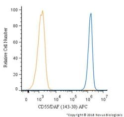
Antigen: CD55/DAF
Concentration: 1.0 mg/mL
Applications: Flow Cytometry, Immunohistochemistry (Frozen), Immunofluorescence, CyTOF
Conjugate: Unconjugated
Host Species: Mouse
Research Discipline: Immunology
Formulation: PBS with No Preservative
Gene ID (Entrez): 1604
Immunogen: PHA stimulated human PBL
Primary or Secondary: Primary
Content And Storage: Store at 4C short term. Aliquot and store at -20C long term. Avoid freeze-thaw cycles.
Molecular Weight of Antigen: 70 kDa
Clone: 143-30
Dilution: Flow Cytometry : 0.5 - 1 ug/million cells in 0.1 ml, Immunohistochemistry-Frozen : 0.5 - 1.0 ug/ml, Immunofluorescence : 0.5 - 1.0 ug/ml, CyTOF-ready
Classification: Monoclonal
Form: Purified
Regulatory Status: RUO
Target Species: Human
Gene Alias: CD55 antigen, CD55 molecule, decay accelerating factor for complement (Cromer blood group), CRdecay accelerating factor for complement (CD55, Cromer blood group system), CROMDAFcomplement decay-accelerating factor, decay accelerating factor for complement, TC
Gene Symbols: CD55
Isotype: IgG1 κ
Purification Method: Protein A or G purified
Test Specificity: Recognizes a single chain glycoprotein of 70kDa, identified as CD55 (also known as decay accelerating factor, DAF). CD55/DAF is widely expressed on cells throughout the body including leukocytes, erythrocytes, epithelium, endothelium, and fibroblasts. It is a Glycosyl phosphatidylinositol anchored (GPI-anchored) member of the membrane bound complement regulatory proteins that inhibit autologous complement cascade activation. It prevents the amplification steps of the complement cascade by interfering with the assembly of the C3-convertases, C4b2a and C3bBb, and the C5-convertase, C4b2a3b and C3bBb3b. CD55 also serves as receptor for CD97 and for echovirus and Coxsackie B virus. The MAb 143-30 can be used as marker for paroxysmal nocturnal hemoglobinuria (PNH).