HLA DR/DP Antibody (Bra-14) - Azide and BSA Free, Novus Biologicals™
Mouse Monoclonal Antibody
Manufacturer: Fischer Scientific
The price for this product is unavailable. Please request a quote
Antigen
HLA DR/DP
Concentration
1.0 mg/mL
Applications
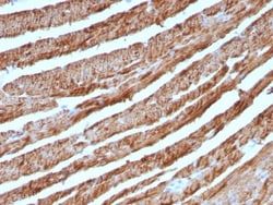
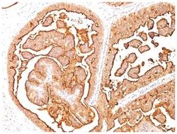
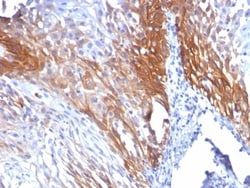
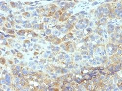
Flow Cytometry, Immunohistochemistry (Paraffin), Immunofluorescence, CyTOF
Conjugate
Unconjugated
Host Species
Mouse
Research Discipline
Adaptive Immunity, Cell Biology, Diabetes Research, Immunology
Formulation
PBS with No Preservative
Gene ID (Entrez)
3122
Immunogen
Human REH cells
Primary or Secondary
Primary
Content And Storage
Store at 4C short term. Aliquot and store at -20C long term. Avoid freeze-thaw cycles.
Clone
Bra-14
Dilution
Flow Cytometry : 0.5 - 1 ug/million cells in 0.1 ml, Immunohistochemistry-Paraffin : 0.5 - 1.0 ug/ml, Immunofluorescence : 0.5 - 1.0 ug/ml, CyTOF-ready
Classification
Monoclonal
Form
Purified
Regulatory Status
RUO
Target Species
Human
Gene Alias
FLJ51114, histocompatibility antigen HLA-DR alpha, HLA class II histocompatibility antigen, DR alpha chain, HLA-DRA1, major histocompatibility complex, class II, DR alpha, MHC cell surface glycoprotein, MHC class II antigen DRA, MLRW
Gene Symbols
HLA-DRA
Isotype
IgG3 κ
Purification Method
Protein A or G purified
Test Specificity
Reacts with a common epitope of human major histocompatibility (MHC) class II antigens, HLA-DR and DP. Human MHC class II antigens are transmembrane glycoproteins composed of an chain (36kDa) and a chain (27kDa). They are expressed primarily on antigen presenting cells such as B lymphocytes, monocytes, macrophages, and thymic epithelial cells and are also present on activated T lymphocytes. Human MHC class II genes are located in the HLA-D region that encodes at least six and ten chain genes. Three loci, DR, DQ and DP, encode the major expressed products of the human class II region. The human MHC class II molecules bind intracellularly processed peptides and present them to T-helper cells. They, therefore, have a critical role in the initiation of the immune response. It has been shown that some autoimmune diseases are associated with certain class II alleles.
Related Products

Description
- HLA DR/DP Monoclonal specifically detects HLA DR/DP in Human samples
- It is validated for Flow Cytometry, Immunohistochemistry, Immunocytochemistry/Immunofluorescence, Immunohistochemistry-Paraffin, Immunofluorescence, CyTOF-ready.
Compare Similar Items
Show Difference

Antigen: HLA DR/DP
Concentration: 1.0 mg/mL
Applications: Flow Cytometry, Immunohistochemistry (Paraffin), Immunofluorescence, CyTOF
Conjugate: Unconjugated
Host Species: Mouse
Research Discipline: Adaptive Immunity, Cell Biology, Diabetes Research, Immunology
Formulation: PBS with No Preservative
Gene ID (Entrez): 3122
Immunogen: Human REH cells
Primary or Secondary: Primary
Content And Storage: Store at 4C short term. Aliquot and store at -20C long term. Avoid freeze-thaw cycles.
Clone: Bra-14
Dilution: Flow Cytometry : 0.5 - 1 ug/million cells in 0.1 ml, Immunohistochemistry-Paraffin : 0.5 - 1.0 ug/ml, Immunofluorescence : 0.5 - 1.0 ug/ml, CyTOF-ready
Classification: Monoclonal
Form: Purified
Regulatory Status: RUO
Target Species: Human
Gene Alias: FLJ51114, histocompatibility antigen HLA-DR alpha, HLA class II histocompatibility antigen, DR alpha chain, HLA-DRA1, major histocompatibility complex, class II, DR alpha, MHC cell surface glycoprotein, MHC class II antigen DRA, MLRW
Gene Symbols: HLA-DRA
Isotype: IgG3 κ
Purification Method: Protein A or G purified
Test Specificity: Reacts with a common epitope of human major histocompatibility (MHC) class II antigens, HLA-DR and DP. Human MHC class II antigens are transmembrane glycoproteins composed of an chain (36kDa) and a chain (27kDa). They are expressed primarily on antigen presenting cells such as B lymphocytes, monocytes, macrophages, and thymic epithelial cells and are also present on activated T lymphocytes. Human MHC class II genes are located in the HLA-D region that encodes at least six and ten chain genes. Three loci, DR, DQ and DP, encode the major expressed products of the human class II region. The human MHC class II molecules bind intracellularly processed peptides and present them to T-helper cells. They, therefore, have a critical role in the initiation of the immune response. It has been shown that some autoimmune diseases are associated with certain class II alleles.
Antigen: SHBG
Concentration: 1.0 mg/mL
Applications: Flow Cytometry, Immunohistochemistry (Paraffin), Immunofluorescence, CyTOF
Conjugate: Unconjugated
Host Species: Mouse
Research Discipline: Cancer, Vision
Formulation: PBS with No Preservative
Gene ID (Entrez): 6462
Immunogen: Recombinant full-length human SHBG protein
Primary or Secondary: Primary
Content And Storage: Store at 4C short term. Aliquot and store at -20C long term. Avoid freeze-thaw cycles.
Clone: SHBG/245
Dilution: Flow Cytometry : 0.5 - 1 ug/million cells in 0.1 ml, Immunohistochemistry-Paraffin : 1 - 2 ug/ml, Immunofluorescence : 0.5 - 1.0 ug/ml, CyTOF-ready
Classification: Monoclonal
Form: Purified
Regulatory Status: RUO
Target Species: Human
Gene Alias: ABPTestosterone-estrogen-binding globulin, androgen binding protein, MGC126834, MGC138391, SBP, sex hormone-binding globulin, Sex steroid-binding protein, TeBG, Testis-specific androgen-binding protein, testosterone-binding beta-globulin, Testosterone-estradiol-binding globulin
Gene Symbols: SHBG
Isotype: IgG1
Purification Method: Protein A or G purified
Test Specificity: Recognizes a protein of 45kDa, identified as SHBG. It functions as an androgen transport protein, but may also be involved in receptor-mediated processes. Each dimer binds one molecule of steroid. It is specific for 5-alpha-dihydrotestosterone, testosterone, and 17-beta-estradiol. SHBG regulates the plasma metabolic clearance rate of steroid hormones by controlling their plasma concentration. In testis, it is synthesized by the Sertoli cells, secreted into the lumen of the seminiferous tubule and transported to the epididymis.