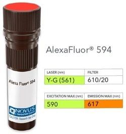
NB005620

Bax Antibody (2D2), DyLight 594, Novus Biologicals™

Manufacturer: Novus Biologicals

Select a Size

Pack Size SKU Availability Price
Each of 1 NB005620-Each-of-1 In Stock ₹ 56,298.48

NB005620 - Each of 1

₹ 56,298.48

In Stock

Quantity

1

Base Price: ₹ 56,298.48

GST (18%): ₹ 10,133.726

Total Price: ₹ 66,432.206

Antigen

Bax

Classification

Monoclonal

Conjugate

DyLight 594

Formulation

50mM Sodium Borate with 0.05% Sodium Azide

Gene Symbols

BAX

Immunogen

A synthetic peptide, aa 3-16 (Cys-GSGEQPRGGGPTSS) of human bax protein. (Uniprot: Q07812)

Quantity

0.1 mL

Research Discipline

Apoptosis, Cancer, Core ESC Like Genes, Stem Cell Markers, Tumor Suppressors

Test Specificity

Recognizes a protein of 21kDa, identified as the Bax protein. This monoclonal antibody is highly specific to Bax and shows no cross-reaction with Bcl-2 or Bcl-X protein. Bcl-2 blocks cell death following a variety of stimuli. Bax has extensive amino acid homology with Bcl-2 and it homodimerizes and forms heterodimers with Bcl-2. Overexpression of Bax accelerates apoptotic death induced by cytokine deprivation in an IL-3 dependent cell line, and Bax also counters the death repressor activity of Bcl-2.

Content And Storage

Store at 4°C in the dark.

Applications

Flow Cytometry, ELISA, Immunocytochemistry, Immunofluorescence, Immunohistochemistry (Paraffin)

Clone

2D2

Dilution

Flow Cytometry, ELISA, Immunocytochemistry/Immunofluorescence, Immunohistochemistry-Paraffin

Gene Alias

apoptosis regulator BAX, BCL2-associated X protein, Bcl2-L-4, BCL2L4bcl2-L-4, Bcl-2-like protein 4

Host Species

Mouse

Purification Method

Protein A or G purified

Regulatory Status

RUO

Primary or Secondary

Primary

Target Species

Human, Monkey, Mouse (Negative), Rat (Negative)

Isotype

IgG1 κ

Related Products

Img

Novus Biologicals

NB005722

--

Img

Novus Biologicals

NB005794

--

Img

Novus Biologicals

NB005792

--

Img

Novus Biologicals

NB005793

--

Img

Novus Biologicals

NB008424

--

Img

Novus Biologicals

NB008284

--

Img

Novus Biologicals

NB008421

--

Img

Novus Biologicals

NB008419

--

Description

  • Bax Monoclonal specifically detects Bax in Human, Monkey, Mouse (Negative), Rat (Negative) samples
  • It is validated for Western Blot, Flow Cytometry, Immunocytochemistry/Immunofluorescence, Immunohistochemistry-Paraffin.

Compare Similar Items

Show Difference

Img

Novus Biologicals

NB005620

--


Antigen:
Bax

Classification:
Monoclonal

Conjugate:
DyLight 594

Formulation:
50mM Sodium Borate with 0.05% Sodium Azide

Gene Symbols:
BAX

Immunogen:
A synthetic peptide, aa 3-16 (Cys-GSGEQPRGGGPTSS) of human bax protein. (Uniprot: Q07812)

Quantity:
0.1 mL

Research Discipline:
Apoptosis, Cancer, Core ESC Like Genes, Stem Cell Markers, Tumor Suppressors

Test Specificity:
Recognizes a protein of 21kDa, identified as the Bax protein. This monoclonal antibody is highly specific to Bax and shows no cross-reaction with Bcl-2 or Bcl-X protein. Bcl-2 blocks cell death following a variety of stimuli. Bax has extensive amino acid homology with Bcl-2 and it homodimerizes and forms heterodimers with Bcl-2. Overexpression of Bax accelerates apoptotic death induced by cytokine deprivation in an IL-3 dependent cell line, and Bax also counters the death repressor activity of Bcl-2.

Content And Storage:
Store at 4°C in the dark.

Applications:
Flow Cytometry, ELISA, Immunocytochemistry, Immunofluorescence, Immunohistochemistry (Paraffin)

Clone:
2D2

Dilution:
Flow Cytometry, ELISA, Immunocytochemistry/Immunofluorescence, Immunohistochemistry-Paraffin

Gene Alias:
apoptosis regulator BAX, BCL2-associated X protein, Bcl2-L-4, BCL2L4bcl2-L-4, Bcl-2-like protein 4

Host Species:
Mouse

Purification Method:
Protein A or G purified

Regulatory Status:
RUO

Primary or Secondary:
Primary

Target Species:
Human, Monkey, Mouse (Negative), Rat (Negative)

Isotype:
IgG1 κ

Img

Novus Biologicals

NB005621

--


Antigen:
MAGE 1

Classification:
Monoclonal

Conjugate:
DyLight 594

Formulation:
50mM Sodium Borate with 0.05% Sodium Azide

Gene Symbols:
MAGEA1

Immunogen:
Recombinant full-length human MAGE 1 protein (Uniprot: P43355)

Quantity:
0.1 mL

Research Discipline:
Apoptosis, Cancer, Melanoma Cell Markers, Tumor Suppressors

Test Specificity:
Recognizes a protein of 42-46kDa, identified as MAGE-1. This monoclonal antibody does not cross-react with MAGE-2, -3, -4, -6 -9, -10, -or -12 protein. Human malignant neoplasms carry rejection antigens that are recognized by the patients' autologous, tumor directed and specific, cytolytic, CD8+ T lymphocyte clones (CTL). The MAGE family of genes codes an important group of antigens. It was identified that melanomas and primary glial brain tumors express common melanoma associated antigens (MAAs). Because MAGE-1 is expressed on a significant proportion of human neoplasms of various histological types (melanoma, brain tumors of glial origin, neuroblastoma, non-small cell lung cancer, breast, gastric, colorectal, ovarian, renal cell carcinomas) and not on normal tissues, the encoded antigen may serve as a marker of early detection and target for cancer immunotherapy.

Content And Storage:
Store at 4°C in the dark.

Applications:
ELISA

Clone:
MA454

Dilution:
ELISA

Gene Alias:
Antigen MZ2-E, Cancer/testis antigen 1.1, cancer/testis antigen family 1, member 1, CT1.1melanoma antigen family A 1, MAGE-1 antigen, MAGE1A, MAGE1melanoma antigen MAGE-1, melanoma antigen family A, 1 (directs expression of antigen MZ2-E), melanoma-associated antigen 1, melanoma-associated antigen MZ2-E, MGC9326

Host Species:
Mouse

Purification Method:
Protein A or G purified

Regulatory Status:
RUO

Primary or Secondary:
Primary

Target Species:
Human, Rat, Canine

Isotype:
IgG1 κ

Img

Novus Biologicals

NB005622

--


Antigen:
MAGE 1

Classification:
Monoclonal

Conjugate:
FITC

Formulation:
PBS with 0.05% Sodium Azide

Gene Symbols:
MAGEA1

Immunogen:
Recombinant full-length human MAGE 1 protein (Uniprot: P43355)

Quantity:
0.1 mL

Research Discipline:
Apoptosis, Cancer, Melanoma Cell Markers, Tumor Suppressors

Test Specificity:
Recognizes a protein of 42-46kDa, identified as MAGE-1. This monoclonal antibody does not cross-react with MAGE-2, -3, -4, -6 -9, -10, -or -12 protein. Human malignant neoplasms carry rejection antigens that are recognized by the patients' autologous, tumor directed and specific, cytolytic, CD8+ T lymphocyte clones (CTL). The MAGE family of genes codes an important group of antigens. It was identified that melanomas and primary glial brain tumors express common melanoma associated antigens (MAAs). Because MAGE-1 is expressed on a significant proportion of human neoplasms of various histological types (melanoma, brain tumors of glial origin, neuroblastoma, non-small cell lung cancer, breast, gastric, colorectal, ovarian, renal cell carcinomas) and not on normal tissues, the encoded antigen may serve as a marker of early detection and target for cancer immunotherapy.

Content And Storage:
Store at 4°C in the dark.

Applications:
ELISA

Clone:
MA454

Dilution:
ELISA

Gene Alias:
Antigen MZ2-E, Cancer/testis antigen 1.1, cancer/testis antigen family 1, member 1, CT1.1melanoma antigen family A 1, MAGE-1 antigen, MAGE1A, MAGE1melanoma antigen MAGE-1, melanoma antigen family A, 1 (directs expression of antigen MZ2-E), melanoma-associated antigen 1, melanoma-associated antigen MZ2-E, MGC9326

Host Species:
Mouse

Purification Method:
Protein A or G purified

Regulatory Status:
RUO

Primary or Secondary:
Primary

Target Species:
Human, Rat, Canine

Isotype:
IgG1 κ

Img

Novus Biologicals

NB005625

--


Antigen:
TIMP-3

Classification:
Polyclonal

Conjugate:
Alexa Fluor 532

Formulation:
50mM Sodium Borate with 0.05% Sodium Azide

Gene Symbols:
TIMP3

Immunogen:
A portion of amino acids 175-211 of human TIMP3 was used as immunogen for this antibody.

Quantity:
0.1 mL

Research Discipline:
Angiogenesis, Apoptosis, Cancer, Hypoxia, Metastasis, Vision

Test Specificity:
TIMP3 (tissue inhibitor of metalloproteinases 3), along with family members TIMP1, TIMP2, and TIMP4, are inhibitors of the matrix metalloproteinases (MMPs), a group of peptidases involved in degradation of the extracellular matrix (ECM). An imbalance between MMPs and the associated TIMPs may play a significant role in the invasive phenotype of malignant tumors. TIMP s inhibit the proteolytic invasiveness of tumor cells and normal placental trophoblast cells. TIMP-3 may be involved in regulating trophoblastic invasion of the uterus as well as in regulating remodeling of the extracellular matrix during the folding of epithelia, and in the formation, branching and expansion of epithelial tubes.

Content And Storage:
Store at 4°C in the dark.

Applications:
Western Blot, Flow Cytometry, Immunohistochemistry, Immunocytochemistry, Immunofluorescence, Immunohistochemistry (Paraffin)

Clone:
__

Dilution:
Western Blot, Flow Cytometry, Immunohistochemistry, Immunocytochemistry/Immunofluorescence, Immunohistochemistry-Paraffin

Gene Alias:
HSMRK222, K222, K222TA2, metalloproteinase inhibitor 3, MIG-5 protein, Protein MIG-5, pseudoinflammatory), SFD, TIMP metallopeptidase inhibitor 3, TIMP-3, tissue inhibitor of metalloproteinase 3 (Sorsby fundus dystrophy, Tissue inhibitor of metalloproteinases 3

Host Species:
Rabbit

Purification Method:
Protein A purified

Regulatory Status:
RUO

Primary or Secondary:
Primary

Target Species:
Human, Bovine, Canine, Equine, Sheep

Isotype:
IgG