NB005794

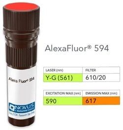
Bcl-2 Antibody (100/D5), DyLight 594, Novus Biologicals™

Manufacturer: Novus Biologicals

Select a Size

Pack Size SKU Availability Price
Each of 1 NB005794-Each-of-1 In Stock ₹ 56,298.48

NB005794 - Each of 1

₹ 56,298.48

In Stock

Quantity

1

Base Price: ₹ 56,298.48

GST (18%): ₹ 10,133.726

Total Price: ₹ 66,432.206

Antigen

Bcl-2

Classification

Monoclonal

Conjugate

DyLight 594

Formulation

50mM Sodium Borate with 0.05% Sodium Azide

Gene Symbols

BCL2

Immunogen

A synthetic peptide, aa41-54 (GAAPAPGIFSSQPG-Cys) of human Bcl-2 protein. (Uniprot: P10415 )

Quantity

0.1 mL

Research Discipline

Apoptosis, Autophagy, Biologically Active Proteins, Cancer, Cellular Markers, Mitochondrial Mediated Pathway, Mitophagy, Oncogenes, Phospho Specific, Tumor Suppressors

Test Specificity

This antibody recognizes a protein of 25-26kDa, identified as the Bcl-2 lpha oncoprotein. It shows no cross-reaction with Bcl-x or Bax protein. Expression of Bcl-2 lpha oncoprotein inhibits the programmed cell death (apoptosis). In most follicular lymphomas, neoplastic germinal centers express high levels of Bcl-2 lpha protein, whereas the normal or hyperplastic germinal centers are negative. Consequently, this antibody is valuable when distinguishing between reactive and neoplastic follicular proliferation in lymph node biopsies. It may also be used in distinguishing between those follicular lymphomas that express Bcl-2 protein and the small number in which the neoplastic cells are Bcl-2 negative.

Content And Storage

Store at 4°C in the dark.

Applications

Western Blot, ELISA, Immunohistochemistry, Immunocytochemistry, Immunofluorescence, Immunohistochemistry (Paraffin)

Clone

100/D5

Dilution

Western Blot, ELISA, Immunohistochemistry, Immunocytochemistry/Immunofluorescence, Immunohistochemistry-Paraffin, Immunohistochemistry-Frozen

Gene Alias

apoptosis regulator Bcl-2, B-cell CLL/lymphoma 2, Bcl-2

Host Species

Mouse

Purification Method

Protein A or G purified

Regulatory Status

RUO

Primary or Secondary

Primary

Target Species

Human, Mouse (Negative), Rat (Negative)

Isotype

IgG1 κ

Related Products

Img

Novus Biologicals

NB005792

--

Img

Novus Biologicals

NB005793

--

Img

Novus Biologicals

NB005620

--

Img

Novus Biologicals

NB008292

--

Img

Novus Biologicals

NB008287

--

Img

Novus Biologicals

NB005719

--

Img

Novus Biologicals

NB005750

--

Img

Novus Biologicals

NB005717

--

Description

  • Bcl-2 Monoclonal specifically detects Bcl-2 in Human, Mouse (Negative), Rat (Negative) samples
  • It is validated for Western Blot, Immunohistochemistry, Immunocytochemistry/Immunofluorescence, Immunohistochemistry-Paraffin, Immunohistochemistry-Frozen.

Compare Similar Items

Show Difference

Img

Novus Biologicals

NB005794

--


Antigen:
Bcl-2

Classification:
Monoclonal

Conjugate:
DyLight 594

Formulation:
50mM Sodium Borate with 0.05% Sodium Azide

Gene Symbols:
BCL2

Immunogen:
A synthetic peptide, aa41-54 (GAAPAPGIFSSQPG-Cys) of human Bcl-2 protein. (Uniprot: P10415 )

Quantity:
0.1 mL

Research Discipline:
Apoptosis, Autophagy, Biologically Active Proteins, Cancer, Cellular Markers, Mitochondrial Mediated Pathway, Mitophagy, Oncogenes, Phospho Specific, Tumor Suppressors

Test Specificity:
This antibody recognizes a protein of 25-26kDa, identified as the Bcl-2 lpha oncoprotein. It shows no cross-reaction with Bcl-x or Bax protein. Expression of Bcl-2 lpha oncoprotein inhibits the programmed cell death (apoptosis). In most follicular lymphomas, neoplastic germinal centers express high levels of Bcl-2 lpha protein, whereas the normal or hyperplastic germinal centers are negative. Consequently, this antibody is valuable when distinguishing between reactive and neoplastic follicular proliferation in lymph node biopsies. It may also be used in distinguishing between those follicular lymphomas that express Bcl-2 protein and the small number in which the neoplastic cells are Bcl-2 negative.

Content And Storage:
Store at 4°C in the dark.

Applications:
Western Blot, ELISA, Immunohistochemistry, Immunocytochemistry, Immunofluorescence, Immunohistochemistry (Paraffin)

Clone:
100/D5

Dilution:
Western Blot, ELISA, Immunohistochemistry, Immunocytochemistry/Immunofluorescence, Immunohistochemistry-Paraffin, Immunohistochemistry-Frozen

Gene Alias:
apoptosis regulator Bcl-2, B-cell CLL/lymphoma 2, Bcl-2

Host Species:
Mouse

Purification Method:
Protein A or G purified

Regulatory Status:
RUO

Primary or Secondary:
Primary

Target Species:
Human, Mouse (Negative), Rat (Negative)

Isotype:
IgG1 κ

Img

Novus Biologicals

NB005795

--


Antigen:
ER alpha/NR3A1

Classification:
Monoclonal

Conjugate:
FITC

Formulation:
PBS with 0.05% Sodium Azide

Gene Symbols:
ESR1

Immunogen:
Recombinant fragment (around aa 2-185) of human ER alpha/NR3A1 protein (exact sequence is proprietary) (Uniprot: P03372)

Quantity:
0.1 mL

Research Discipline:
Angiogenesis, Breast Cancer, Cancer, Cell Biology, Cell Cycle and Replication, Dendritic Cell Markers, GPCR, Neuroscience, Phospho Specific, Signal Transduction, Transcription Factors and Regulators

Test Specificity:
This monoclonal antibody is specific to ER alpha and shows minimal cross-reaction with other members of the family. ER is an important regulator of growth and differentiation in the mammary gland. Presence of ER in breast tumors indicates an increased likelihood of response to anti-estrogen (e.g. tamoxifen) therapy. It strongly stains nuclei of epithelial cells in breast carcinomas.

Content And Storage:
Store at 4°C in the dark.

Applications:
Flow Cytometry, Immunohistochemistry, Immunohistochemistry (Paraffin), Immunohistochemistry (Frozen)

Clone:
ER506

Dilution:
Flow Cytometry, Immunohistochemistry, Immunohistochemistry-Paraffin, Immunohistochemistry-Frozen, Flow (Intracellular)

Gene Alias:
ER, ER alpha, Era, ER-alpha, ESRESRA, Estradiol receptor, estrogen receptor, estrogen receptor 1, estrogen receptor alpha, estrogen receptor alpha delta 3*4,56,7*/819-2 isoform, estrogen receptor alpha delta 4 +49 isoform, estrogen receptor alpha delta 4*5,6,7*/654 isoform, NR3A1DKFZp686N23123, Nuclear receptor subfamily 3 group A member 1

Host Species:
Mouse

Purification Method:
Protein A or G purified

Regulatory Status:
RUO

Primary or Secondary:
Primary

Target Species:
Human

Isotype:
IgG1 κ

Img

Novus Biologicals

NB005797

--


Antigen:
Progesterone R/NR3C3

Classification:
Monoclonal

Conjugate:
FITC

Formulation:
PBS with 0.05% Sodium Azide

Gene Symbols:
PGR

Immunogen:
Recombinant full-length human Progesterone R/NR3C3 protein (PGR) (Uniprot: P06401 )

Quantity:
0.1 mL

Research Discipline:
Breast Cancer, Cancer, Phospho Specific

Test Specificity:
This monoclonal antibody is specific to progesterone receptor and shows minimal cross-reaction with other members of the family. Progesterone receptor is expressed as two major isoforms, PR-A (81kDa) and PR-B (116kDa). Expression of PgR has been suggested to reflect a intact estrogen regulatory machinery and therefore, predict better clinical response to endocrine therapy than ER alone. It is excellent for immunohistochemical staining of formalin/paraffin tissues.

Content And Storage:
Store at 4°C in the dark.

Applications:
Western Blot, Immunohistochemistry, Immunohistochemistry (Paraffin), Immunocytochemistry, Immunofluorescence

Clone:
PR484

Dilution:
Western Blot, Immunohistochemistry, Immunohistochemistry-Paraffin

Gene Alias:
NR3C3PRNuclear receptor subfamily 3 group C member 3, progesterone receptor

Host Species:
Mouse

Purification Method:
Protein A or G purified

Regulatory Status:
RUO

Primary or Secondary:
Primary

Target Species:
Human

Isotype:
IgG1 κ

Img

Novus Biologicals

NB005798

--


Antigen:
Chorionic Gonadotropin beta Chain (hCG beta)

Classification:
Monoclonal

Conjugate:
Alexa Fluor 350

Formulation:
50mM Sodium Borate with 0.05% Sodium Azide

Gene Symbols:
CGB3

Immunogen:
Recombinant Chorionic Gonadotropin beta Chain (hCG beta) protein (Uniprot: P01233)

Quantity:
0.1 mL

Research Discipline:
Cancer

Test Specificity:
This monoclonal antibody reacts with a protein of 22kDa, identified as beta sub-unit of HCG. It does not cross react with the alpha sub-unit. HCG is a glycoprotein, which is secreted in large quantities by normal trophoblasts. It is present only in trace amounts in non-pregnant urine and sera but rises sharply during pregnancy. HCG is composed of two non-identical, non-covalently linked polypeptide chains designated as the subunit is identical to that of thyroid stimulating hormone (TSH) follicle stimulating hormone (FSH), and luteinizing hormone (LH). hCG monoclonal antibody detects cells and tumors of trophoblastic origin such as choriocarcinoma. Large cell carcinoma and adenocarcinoma of the lung demonstrate anti-hCG positivity in 90% and 60% of cases respectively. 20% of lung squamous cell carcinomas are positive. hCG expression by non-trophoblastic tumors may indicate aggressive behavior.

Content And Storage:
Store at 4°C in the dark.

Applications:
Western Blot, ELISA, Immunohistochemistry, Immunohistochemistry (Paraffin), Immunohistochemistry (Frozen)

Clone:
SPM105

Dilution:
Western Blot, ELISA, Immunohistochemistry, Immunohistochemistry-Paraffin, Immunohistochemistry-Frozen

Gene Alias:
CGB3choriogonadotropin subunit beta, CGB5, CGB7, CGB8, CG-beta, Chorionic gonadotrophin chain beta, chorionic gonadotropin beta 3 subunit, chorionic gonadotropin beta chain, chorionic gonadotropin beta subunit, chorionic gonadotropin, beta polypeptide, hCGB

Host Species:
Mouse

Purification Method:
Protein A or G purified

Regulatory Status:
RUO

Primary or Secondary:
Primary

Target Species:
Human

Isotype:
IgG1 κ