NB005850

Cytokeratin 8 Antibody (SPM192), FITC, Novus Biologicals™

Manufacturer: Novus Biologicals

Select a Size

Pack Size SKU Availability Price
Each of 1 NB005850-Each-of-1 In Stock ₹ 55,271.76

NB005850 - Each of 1

₹ 55,271.76

In Stock

Quantity

1

Base Price: ₹ 55,271.76

GST (18%): ₹ 9,948.917

Total Price: ₹ 65,220.677

Antigen

Cytokeratin 8

Classification

Monoclonal

Conjugate

FITC

Formulation

PBS with 0.05% Sodium Azide

Gene Symbols

KRT8

Immunogen

Keratin preparation from a human carcinoma (Uniprot: P05787)

Quantity

0.1 mL

Research Discipline

Cancer, Cell Biology, Cellular Markers, Phospho Specific

Test Specificity

Epitope of this monoclonal antibody is located between aa343-357. Cytokeratin 8 (CK8) belongs to the type II (or B or basic) subfamily of high molecular weight cytokeratins and exists in combination with cytokeratin 18 (CK18). CK8 is primarily found in the non-squamous epithelia and is present in majority of adenocarcinomas and ductal carcinomas. It is absent in squamous cell carcinomas. Hepatocellular carcinomas are defined by the use of antibodies that recognize only cytokeratin 8 and 18. CK8 exists on several types of normal and neoplastic epithelia, including many ductal and glandular epithelia such as colon, stomach, small intestine, trachea, and esophagus as well as in transitional epithelium. Anti-CK8 does not react with skeletal muscle or nerve cells. Epithelioid sarcoma, chordoma, and adamantinoma show strong positivity corresponding to that of simple epithelia (with antibodies against CK8, CK18 and CK19). Reportedly, anti-CK8 is useful for the differentiation of lobular () carcinoma of the breast.

Content And Storage

Store at 4°C in the dark.

Applications

Flow Cytometry, ELISA, Immunohistochemistry, Immunocytochemistry, Immunofluorescence, Immunohistochemistry (Paraffin)

Clone

SPM192

Dilution

Flow Cytometry, ELISA, Immunohistochemistry, Immunocytochemistry/Immunofluorescence, Immunohistochemistry-Paraffin, Immunohistochemistry-Frozen

Gene Alias

CARD2, CK8, CK-8, CYK8, Cytokeratin-8, K2C8, K8, keratin 8, keratin, type II cytoskeletal 8, keratin-8, KO, Type-II keratin Kb8

Host Species

Mouse

Purification Method

Protein A or G purified

Regulatory Status

RUO

Primary or Secondary

Primary

Target Species

Human, Rat (Negative)

Isotype

IgG1 κ

Related Products

Img

Novus Biologicals

NB005845

--

Img

Novus Biologicals

NB005847

--

Img

Novus Biologicals

NB005848

--

Img

Novus Biologicals

NB005846

--

Img

Novus Biologicals

NB006262

--

Img

Novus Biologicals

NB005950

--

Img

Novus Biologicals

NB005945

--

Img

Novus Biologicals

NB005947

--

Description

  • Cytokeratin 8 Monoclonal specifically detects Cytokeratin 8 in Human, Rat (Negative) samples
  • It is validated for Flow Cytometry, Immunohistochemistry, Immunocytochemistry/Immunofluorescence, Immunohistochemistry-Paraffin.

Compare Similar Items

Show Difference

Img

Novus Biologicals

NB005850

--


Antigen:
Cytokeratin 8

Classification:
Monoclonal

Conjugate:
FITC

Formulation:
PBS with 0.05% Sodium Azide

Gene Symbols:
KRT8

Immunogen:
Keratin preparation from a human carcinoma (Uniprot: P05787)

Quantity:
0.1 mL

Research Discipline:
Cancer, Cell Biology, Cellular Markers, Phospho Specific

Test Specificity:
Epitope of this monoclonal antibody is located between aa343-357. Cytokeratin 8 (CK8) belongs to the type II (or B or basic) subfamily of high molecular weight cytokeratins and exists in combination with cytokeratin 18 (CK18). CK8 is primarily found in the non-squamous epithelia and is present in majority of adenocarcinomas and ductal carcinomas. It is absent in squamous cell carcinomas. Hepatocellular carcinomas are defined by the use of antibodies that recognize only cytokeratin 8 and 18. CK8 exists on several types of normal and neoplastic epithelia, including many ductal and glandular epithelia such as colon, stomach, small intestine, trachea, and esophagus as well as in transitional epithelium. Anti-CK8 does not react with skeletal muscle or nerve cells. Epithelioid sarcoma, chordoma, and adamantinoma show strong positivity corresponding to that of simple epithelia (with antibodies against CK8, CK18 and CK19). Reportedly, anti-CK8 is useful for the differentiation of lobular () carcinoma of the breast.

Content And Storage:
Store at 4°C in the dark.

Applications:
Flow Cytometry, ELISA, Immunohistochemistry, Immunocytochemistry, Immunofluorescence, Immunohistochemistry (Paraffin)

Clone:
SPM192

Dilution:
Flow Cytometry, ELISA, Immunohistochemistry, Immunocytochemistry/Immunofluorescence, Immunohistochemistry-Paraffin, Immunohistochemistry-Frozen

Gene Alias:
CARD2, CK8, CK-8, CYK8, Cytokeratin-8, K2C8, K8, keratin 8, keratin, type II cytoskeletal 8, keratin-8, KO, Type-II keratin Kb8

Host Species:
Mouse

Purification Method:
Protein A or G purified

Regulatory Status:
RUO

Primary or Secondary:
Primary

Target Species:
Human, Rat (Negative)

Isotype:
IgG1 κ

Img

Novus Biologicals

NB005851

--


Antigen:
GFAP

Classification:
Monoclonal

Conjugate:
Alexa Fluor 350

Formulation:
50mM Sodium Borate with 0.05% Sodium Azide

Gene Symbols:
GFAP

Immunogen:
GFAP isolated from pig spinal cord (Uniprot: P14136)

Quantity:
0.1 mL

Research Discipline:
Astrocyte Markers, Cancer, Cell Biology, Cellular Markers, Chaperone Mediated Autophagy (CMA), Inflammation, Neurodegeneration, Neuroscience, Signal Transduction, Stem Cell Markers

Test Specificity:
This monoclonal antibody recognizes a protein of ∼50kDa which is identified as Glial Fibrillary Acidic Protein (GFAP). It shows no cross-reaction with other intermediate filament proteins. GFAP is specifically found in astroglia. GFAP is a very popular marker for localizing benign astrocyte and neoplastic cells of glial origin in the central nervous system. Antibody to GFAP is useful in differentiating primary gliomas from metastatic lesions in the brain and for documenting astrocytic differentiation in tumors outside the CNS.

Content And Storage:
Store at 4°C in the dark.

Applications:
Western Blot, Flow Cytometry, ELISA, Immunohistochemistry, Immunocytochemistry, Immunofluorescence, Immunohistochemistry (Paraffin)

Clone:
SPM248

Dilution:
Western Blot, Flow Cytometry, ELISA, Immunohistochemistry, Immunocytochemistry/Immunofluorescence, Immunohistochemistry-Paraffin, Immunohistochemistry-Frozen

Gene Alias:
FLJ45472, GFAP astrocytes, glial fibrillary acidic protein

Host Species:
Mouse

Purification Method:
Protein A or G purified

Regulatory Status:
RUO

Primary or Secondary:
Primary

Target Species:
Human, Mouse, Rat, Porcine, Bovine, Chicken, Rabbit

Isotype:
IgG1 κ

Img

Novus Biologicals

NB005854

--


Antigen:
GFAP

Classification:
Monoclonal

Conjugate:
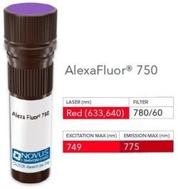
Alexa Fluor 750

Formulation:
50mM Sodium Borate with 0.05% Sodium Azide

Gene Symbols:
GFAP

Immunogen:
GFAP isolated from pig spinal cord (Uniprot: P14136)

Quantity:
0.1 mL

Research Discipline:
Astrocyte Markers, Cancer, Cell Biology, Cellular Markers, Chaperone Mediated Autophagy (CMA), Inflammation, Neurodegeneration, Neuroscience, Signal Transduction, Stem Cell Markers

Test Specificity:
This monoclonal antibody recognizes a protein of ∼50kDa which is identified as Glial Fibrillary Acidic Protein (GFAP). It shows no cross-reaction with other intermediate filament proteins. GFAP is specifically found in astroglia. GFAP is a very popular marker for localizing benign astrocyte and neoplastic cells of glial origin in the central nervous system. Antibody to GFAP is useful in differentiating primary gliomas from metastatic lesions in the brain and for documenting astrocytic differentiation in tumors outside the CNS.

Content And Storage:
Store at 4°C in the dark.

Applications:
Western Blot, Flow Cytometry, ELISA, Immunohistochemistry, Immunocytochemistry, Immunofluorescence, Immunohistochemistry (Paraffin)

Clone:
SPM248

Dilution:
Western Blot, Flow Cytometry, ELISA, Immunohistochemistry, Immunocytochemistry/Immunofluorescence, Immunohistochemistry-Paraffin, Immunohistochemistry-Frozen

Gene Alias:
FLJ45472, GFAP astrocytes, glial fibrillary acidic protein

Host Species:
Mouse

Purification Method:
Protein A or G purified

Regulatory Status:
RUO

Primary or Secondary:
Primary

Target Species:
Human, Mouse, Rat, Porcine, Bovine, Chicken, Rabbit

Isotype:
IgG1 κ

Img

Novus Biologicals

NB005855

--


Antigen:
GFAP

Classification:
Monoclonal

Conjugate:
DyLight 594

Formulation:
50mM Sodium Borate with 0.05% Sodium Azide

Gene Symbols:
GFAP

Immunogen:
GFAP isolated from pig spinal cord (Uniprot: P14136)

Quantity:
0.1 mL

Research Discipline:
Astrocyte Markers, Cancer, Cell Biology, Cellular Markers, Chaperone Mediated Autophagy (CMA), Inflammation, Neurodegeneration, Neuroscience, Signal Transduction, Stem Cell Markers

Test Specificity:
This monoclonal antibody recognizes a protein of ∼50kDa which is identified as Glial Fibrillary Acidic Protein (GFAP). It shows no cross-reaction with other intermediate filament proteins. GFAP is specifically found in astroglia. GFAP is a very popular marker for localizing benign astrocyte and neoplastic cells of glial origin in the central nervous system. Antibody to GFAP is useful in differentiating primary gliomas from metastatic lesions in the brain and for documenting astrocytic differentiation in tumors outside the CNS.

Content And Storage:
Store at 4°C in the dark.

Applications:
Western Blot, Flow Cytometry, ELISA, Immunohistochemistry, Immunocytochemistry, Immunofluorescence, Immunohistochemistry (Paraffin)

Clone:
SPM248

Dilution:
Western Blot, Flow Cytometry, ELISA, Immunohistochemistry, Immunocytochemistry/Immunofluorescence, Immunohistochemistry-Paraffin, Immunohistochemistry-Frozen

Gene Alias:
FLJ45472, GFAP astrocytes, glial fibrillary acidic protein

Host Species:
Mouse

Purification Method:
Protein A or G purified

Regulatory Status:
RUO

Primary or Secondary:
Primary

Target Species:
Human, Mouse, Rat, Porcine, Bovine, Chicken, Rabbit

Isotype:
IgG1 κ