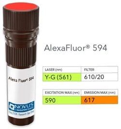
NB005867

Cytokeratin 14 Antibody (SPM263), DyLight 594, Novus Biologicals™

Manufacturer: Novus Biologicals

Select a Size

Pack Size SKU Availability Price
Each of 1 NB005867-Each-of-1 In Stock ₹ 55,271.76

NB005867 - Each of 1

₹ 55,271.76

In Stock

Quantity

1

Base Price: ₹ 55,271.76

GST (18%): ₹ 9,948.917

Total Price: ₹ 65,220.677

Antigen

Cytokeratin 14

Classification

Monoclonal

Conjugate

DyLight 594

Formulation

50mM Sodium Borate with 0.05% Sodium Azide

Gene Symbols

KRT14

Immunogen

A synthetic peptide of 15 amino acid from the C-terminus of human Cytokeratin 14. (Uniprot: P02533)

Quantity

0.1 mL

Research Discipline

Cell Biology, Cellular Markers

Test Specificity

Cytokeratin 14 (CK14) belongs to the type I (or A or acidic) subfamily of low molecular weight keratins and exists in combination with keratin 5 (type II or B or basic). CK14 is found in basal cells of squamous epithelia, some glandular epithelia, myoepithelium, and mesothelial cells. Anti-CK14 is useful in differentiating squamous cell carcinomas from poorly differentiated epithelial tumors. Anti-CK14 is one of the specific basal markers for distinguishing between basal and non-basal subtypes of breast carcinomas. Anti-CK14 is also a good marker for differentiation of intraductal from invasive salivary duct carcinoma by the positive staining of basal cells surrounding the in-situ neoplasm as well as for differentiation of benign prostate from prostate carcinoma. Furthermore, this antibody has been useful in separating oncocytic tumors of the kidney from its renal mimics, and in identifying metaplastic carcinomas of the breast.

Content And Storage

Store at 4°C in the dark.

Applications

Western Blot, Flow Cytometry, ELISA, Immunohistochemistry, Immunocytochemistry, Immunofluorescence, Immunohistochemistry (Paraffin)

Clone

SPM263

Dilution

Western Blot, Flow Cytometry, ELISA, Immunohistochemistry, Immunocytochemistry/Immunofluorescence, Immunohistochemistry-Paraffin, Immunohistochemistry-Frozen

Gene Alias

CK14, CK-14, cytokeratin 14, cytokeratin-14, EBS3, EBS4, K14, keratin 14, keratin 14 (epidermolysis bullosa simplex, Dowling-Meara, Koebner), keratin, type I cytoskeletal 14, Keratin-14, NFJ

Host Species

Mouse

Purification Method

Protein A or G purified

Regulatory Status

RUO

Primary or Secondary

Primary

Target Species

Human, Mouse, Rat

Isotype

IgG3 κ

Related Products

Img

Novus Biologicals

NB005863

--

Img

Novus Biologicals

NB005866

--

Img

Novus Biologicals

NB005868

--

Img

Novus Biologicals

NB006321

--

Img

Novus Biologicals

NB005947

--

Img

Novus Biologicals

NB005847

--

Img

Novus Biologicals

NB005945

--

Img

Novus Biologicals

NB005845

--

Description

  • Cytokeratin 14 Monoclonal specifically detects Cytokeratin 14 in Human, Mouse, Rat samples
  • It is validated for Flow Cytometry, Immunohistochemistry, Immunocytochemistry/Immunofluorescence, Immunohistochemistry-Paraffin, Flow (Intracellular).

Compare Similar Items

Show Difference

Img

Novus Biologicals

NB005867

--


Antigen:
Cytokeratin 14

Classification:
Monoclonal

Conjugate:
DyLight 594

Formulation:
50mM Sodium Borate with 0.05% Sodium Azide

Gene Symbols:
KRT14

Immunogen:
A synthetic peptide of 15 amino acid from the C-terminus of human Cytokeratin 14. (Uniprot: P02533)

Quantity:
0.1 mL

Research Discipline:
Cell Biology, Cellular Markers

Test Specificity:
Cytokeratin 14 (CK14) belongs to the type I (or A or acidic) subfamily of low molecular weight keratins and exists in combination with keratin 5 (type II or B or basic). CK14 is found in basal cells of squamous epithelia, some glandular epithelia, myoepithelium, and mesothelial cells. Anti-CK14 is useful in differentiating squamous cell carcinomas from poorly differentiated epithelial tumors. Anti-CK14 is one of the specific basal markers for distinguishing between basal and non-basal subtypes of breast carcinomas. Anti-CK14 is also a good marker for differentiation of intraductal from invasive salivary duct carcinoma by the positive staining of basal cells surrounding the in-situ neoplasm as well as for differentiation of benign prostate from prostate carcinoma. Furthermore, this antibody has been useful in separating oncocytic tumors of the kidney from its renal mimics, and in identifying metaplastic carcinomas of the breast.

Content And Storage:
Store at 4°C in the dark.

Applications:
Western Blot, Flow Cytometry, ELISA, Immunohistochemistry, Immunocytochemistry, Immunofluorescence, Immunohistochemistry (Paraffin)

Clone:
SPM263

Dilution:
Western Blot, Flow Cytometry, ELISA, Immunohistochemistry, Immunocytochemistry/Immunofluorescence, Immunohistochemistry-Paraffin, Immunohistochemistry-Frozen

Gene Alias:
CK14, CK-14, cytokeratin 14, cytokeratin-14, EBS3, EBS4, K14, keratin 14, keratin 14 (epidermolysis bullosa simplex, Dowling-Meara, Koebner), keratin, type I cytoskeletal 14, Keratin-14, NFJ

Host Species:
Mouse

Purification Method:
Protein A or G purified

Regulatory Status:
RUO

Primary or Secondary:
Primary

Target Species:
Human, Mouse, Rat

Isotype:
IgG3 κ

Img

Novus Biologicals

NB005868

--


Antigen:
Cytokeratin 14

Classification:
Monoclonal

Conjugate:
FITC

Formulation:
PBS with 0.05% Sodium Azide

Gene Symbols:
KRT14

Immunogen:
A synthetic peptide of 15 amino acid from the C-terminus of human Cytokeratin 14. (Uniprot: P02533)

Quantity:
0.1 mL

Research Discipline:
Cell Biology, Cellular Markers

Test Specificity:
Cytokeratin 14 (CK14) belongs to the type I (or A or acidic) subfamily of low molecular weight keratins and exists in combination with keratin 5 (type II or B or basic). CK14 is found in basal cells of squamous epithelia, some glandular epithelia, myoepithelium, and mesothelial cells. Anti-CK14 is useful in differentiating squamous cell carcinomas from poorly differentiated epithelial tumors. Anti-CK14 is one of the specific basal markers for distinguishing between basal and non-basal subtypes of breast carcinomas. Anti-CK14 is also a good marker for differentiation of intraductal from invasive salivary duct carcinoma by the positive staining of basal cells surrounding the in-situ neoplasm as well as for differentiation of benign prostate from prostate carcinoma. Furthermore, this antibody has been useful in separating oncocytic tumors of the kidney from its renal mimics, and in identifying metaplastic carcinomas of the breast.

Content And Storage:
Store at 4°C in the dark.

Applications:
Flow Cytometry, ELISA, Immunohistochemistry, Immunocytochemistry, Immunofluorescence, Immunohistochemistry (Paraffin)

Clone:
SPM263

Dilution:
Flow Cytometry, ELISA, Immunohistochemistry, Immunocytochemistry/Immunofluorescence, Immunohistochemistry-Paraffin, Immunohistochemistry-Frozen

Gene Alias:
CK14, CK-14, cytokeratin 14, cytokeratin-14, EBS3, EBS4, K14, keratin 14, keratin 14 (epidermolysis bullosa simplex, Dowling-Meara, Koebner), keratin, type I cytoskeletal 14, Keratin-14, NFJ

Host Species:
Mouse

Purification Method:
Protein A or G purified

Regulatory Status:
RUO

Primary or Secondary:
Primary

Target Species:
Human, Mouse, Rat

Isotype:
IgG3 κ

Img

Novus Biologicals

NB005870

--


Antigen:
Cytokeratin 18

Classification:
Monoclonal

Conjugate:
Alexa Fluor 532

Formulation:
50mM Sodium Borate with 0.05% Sodium Azide

Gene Symbols:
KRT18

Immunogen:
PMC-42 human breast carcinoma cells

Quantity:
0.1 mL

Research Discipline:
Cancer, Cell Biology, Cellular Markers, Phospho Specific

Test Specificity:
This monoclonal antibody reacts with a wide variety of simple epithelia. It does not react with stratified squamous epithelia. It reacts with epithelial tumors of the gastrointestinal tract, lung, breast, pancreas, ovary, and thyroid. Cytokeratin 18, which belongs to the type A (acidic) subfamily of low molecular weight keratins, exists in combination with cytokeratin 8. It is reported that tissues from gastrointestinal tract are positive for both cytokeratin 8 and 18 but do not contain cytokeratin 14. Tissues from gastrointestinal tract, respiratory tract and urogenital tract, as well as endocrine and exocrine tissues and mesothelial cells are positive for cytokeratin 18.

Content And Storage:
Store at 4°C in the dark.

Applications:
Western Blot, Flow Cytometry, ELISA, Immunohistochemistry, Immunocytochemistry, Immunofluorescence, Immunohistochemistry (Paraffin)

Clone:
SPM265

Dilution:
Western Blot, Flow Cytometry, ELISA, Immunohistochemistry, Immunocytochemistry/Immunofluorescence, Immunohistochemistry-Paraffin, Immunohistochemistry-Frozen

Gene Alias:
Cell proliferation-inducing gene 46 protein, cell proliferation-inducing protein 46, CK-18, CYK18, cytokeratin 18, cytokeratin-18, K18, keratin 18, keratin, type I cytoskeletal 18, keratin-18

Host Species:
Mouse

Purification Method:
Protein A or G purified

Regulatory Status:
RUO

Primary or Secondary:
Primary

Target Species:
Human, Bovine (Negative), Canine (Negative), Hamster (Negative), Mouse (Negative), Porcine (Negative), Rat (Negative)

Isotype:
IgG1 κ

Img

Novus Biologicals

NB005871

--


Antigen:
Cytokeratin 18

Classification:
Monoclonal

Conjugate:
Alexa Fluor 594

Formulation:
50mM Sodium Borate with 0.05% Sodium Azide

Gene Symbols:
KRT18

Immunogen:
PMC-42 human breast carcinoma cells

Quantity:
0.1 mL

Research Discipline:
Cancer, Cell Biology, Cellular Markers, Phospho Specific

Test Specificity:
This monoclonal antibody reacts with a wide variety of simple epithelia. It does not react with stratified squamous epithelia. It reacts with epithelial tumors of the gastrointestinal tract, lung, breast, pancreas, ovary, and thyroid. Cytokeratin 18, which belongs to the type A (acidic) subfamily of low molecular weight keratins, exists in combination with cytokeratin 8. It is reported that tissues from gastrointestinal tract are positive for both cytokeratin 8 and 18 but do not contain cytokeratin 14. Tissues from gastrointestinal tract, respiratory tract and urogenital tract, as well as endocrine and exocrine tissues and mesothelial cells are positive for cytokeratin 18.

Content And Storage:
Store at 4°C in the dark.

Applications:
Western Blot, Flow Cytometry, ELISA, Immunohistochemistry, Immunocytochemistry, Immunofluorescence, Immunohistochemistry (Paraffin)

Clone:
SPM265

Dilution:
Western Blot, Flow Cytometry, ELISA, Immunohistochemistry, Immunocytochemistry/Immunofluorescence, Immunohistochemistry-Paraffin, Immunohistochemistry-Frozen

Gene Alias:
Cell proliferation-inducing gene 46 protein, cell proliferation-inducing protein 46, CK-18, CYK18, cytokeratin 18, cytokeratin-18, K18, keratin 18, keratin, type I cytoskeletal 18, keratin-18

Host Species:
Mouse

Purification Method:
Protein A or G purified

Regulatory Status:
RUO

Primary or Secondary:
Primary

Target Species:
Human, Bovine (Negative), Canine (Negative), Hamster (Negative), Mouse (Negative), Porcine (Negative), Rat (Negative)

Isotype:
IgG1 κ