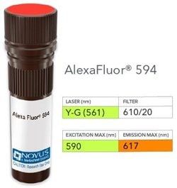
NB006101

alpha-Methylacyl-CoA Racemase/AMACR, Rabbit anti-Human, DyLight 594, Polyclonal, Novus Biologicals™

Manufacturer: Novus Biologicals

Select a Size

Pack Size SKU Availability Price
Each of 1 NB006101-Each-of-1 In Stock ₹ 52,277.16

NB006101 - Each of 1

₹ 52,277.16

In Stock

Quantity

1

Base Price: ₹ 52,277.16

GST (18%): ₹ 9,409.889

Total Price: ₹ 61,687.049

Antigen

alpha-Methylacyl-CoA Racemase/AMACR

Classification

Polyclonal

Dilution

Western Blot, Immunohistochemistry, Immunohistochemistry-Paraffin

Gene Alias

2-methylacyl-CoA racemase, alpha-methylacyl-CoA racemase, CBAS4, EC 5.1.99.4, RACE, RM

Host Species

Rabbit

Purification Method

Protein A purified

Regulatory Status

RUO

Primary or Secondary

Primary

Target Species

Human

Isotype

IgG

Applications

Western Blot, Immunohistochemistry, Immunohistochemistry (Paraffin)

Conjugate

DyLight 594

Formulation

50mM Sodium Borate with 0.05% Sodium Azide

Gene Symbols

AMACR

Immunogen

A synthetic peptide from human AMACR protein

Quantity

0.1 mL

Research Discipline

Lipid and Metabolism, Prostate Cancer, Signal Transduction

Test Specificity

This antibody recognizes a protein of 54kDa, which is identified as AMACR, also known as p504S. It is an enzyme that is involved in bile acid biosynthesis and oxidation of branched-chain fatty acids. AMACR is essential in lipid metabolism. It is expressed in cells of premalignant high-grade prostatic intraepithelial neoplasia (HGPIN) and prostate adenocarcinoma. The majority of the carcinoma cells show a distinct granular cytoplasmic staining reaction. AMACR is present at low or undetectable levels in glandular epithelial cells of normal prostate and benign prostatic hyperplasia. A spotty granular cytoplasmic staining is seen in a few cells of the benign glands. AMACR is expressed in normal liver (hepatocytes), kidney (tubular epithelial cells) and gall bladder (epithelial cells). Expression has also been found in lung (bronchial epithelial cells) and colon (colonic surface epithelium). AMACR expression can also be found in hepatocellular carcinoma and kidney carcinoma. Past studies have also shown that AMACR is expressed in various colon carcinomas (well, moderately and poorly differentiated) and over expressed in prostate carcinoma.

Content And Storage

Store at 4°C in the dark.

Related Products

Img

Novus Biologicals

NB006097

--

Img

Novus Biologicals

NB006104

--

Img

Novus Biologicals

NB006703

--

Img

Novus Biologicals

NB006701

--

Img

Novus Biologicals

NB006446

--

Img

Novus Biologicals

NB006449

--

Img

Novus Biologicals

NB006699

--

Img

Novus Biologicals

NB006702

--

Description

  • alpha-Methylacyl-CoA Racemase/AMACR Polyclonal specifically detects alpha-Methylacyl-CoA Racemase/AMACR in Human samples
  • It is validated for Western Blot, Immunohistochemistry, Immunohistochemistry-Paraffin.

Compare Similar Items

Show Difference

Img

Novus Biologicals

NB006101

--


Antigen:
alpha-Methylacyl-CoA Racemase/AMACR

Classification:
Polyclonal

Dilution:
Western Blot, Immunohistochemistry, Immunohistochemistry-Paraffin

Gene Alias:
2-methylacyl-CoA racemase, alpha-methylacyl-CoA racemase, CBAS4, EC 5.1.99.4, RACE, RM

Host Species:
Rabbit

Purification Method:
Protein A purified

Regulatory Status:
RUO

Primary or Secondary:
Primary

Target Species:
Human

Isotype:
IgG

Applications:
Western Blot, Immunohistochemistry, Immunohistochemistry (Paraffin)

Conjugate:
DyLight 594

Formulation:
50mM Sodium Borate with 0.05% Sodium Azide

Gene Symbols:
AMACR

Immunogen:
A synthetic peptide from human AMACR protein

Quantity:
0.1 mL

Research Discipline:
Lipid and Metabolism, Prostate Cancer, Signal Transduction

Test Specificity:
This antibody recognizes a protein of 54kDa, which is identified as AMACR, also known as p504S. It is an enzyme that is involved in bile acid biosynthesis and oxidation of branched-chain fatty acids. AMACR is essential in lipid metabolism. It is expressed in cells of premalignant high-grade prostatic intraepithelial neoplasia (HGPIN) and prostate adenocarcinoma. The majority of the carcinoma cells show a distinct granular cytoplasmic staining reaction. AMACR is present at low or undetectable levels in glandular epithelial cells of normal prostate and benign prostatic hyperplasia. A spotty granular cytoplasmic staining is seen in a few cells of the benign glands. AMACR is expressed in normal liver (hepatocytes), kidney (tubular epithelial cells) and gall bladder (epithelial cells). Expression has also been found in lung (bronchial epithelial cells) and colon (colonic surface epithelium). AMACR expression can also be found in hepatocellular carcinoma and kidney carcinoma. Past studies have also shown that AMACR is expressed in various colon carcinomas (well, moderately and poorly differentiated) and over expressed in prostate carcinoma.

Content And Storage:
Store at 4°C in the dark.

Img

Novus Biologicals

NB006104

--


Antigen:
alpha-Methylacyl-CoA Racemase/AMACR

Classification:
Polyclonal

Dilution:
Western Blot, Immunohistochemistry, Immunohistochemistry-Paraffin

Gene Alias:
2-methylacyl-CoA racemase, alpha-methylacyl-CoA racemase, CBAS4, EC 5.1.99.4, RACE, RM

Host Species:
Rabbit

Purification Method:
Protein A purified

Regulatory Status:
RUO

Primary or Secondary:
Primary

Target Species:
Human

Isotype:
IgG

Applications:
Western Blot, Immunohistochemistry, Immunohistochemistry (Paraffin)

Conjugate:
Janelia Fluor 646

Formulation:
50mM Sodium Borate with 0.05% Sodium Azide

Gene Symbols:
AMACR

Immunogen:
A synthetic peptide from human AMACR protein

Quantity:
0.1 mL

Research Discipline:
Lipid and Metabolism, Prostate Cancer, Signal Transduction

Test Specificity:
This antibody recognizes a protein of 54kDa, which is identified as AMACR, also known as p504S. It is an enzyme that is involved in bile acid biosynthesis and oxidation of branched-chain fatty acids. AMACR is essential in lipid metabolism. It is expressed in cells of premalignant high-grade prostatic intraepithelial neoplasia (HGPIN) and prostate adenocarcinoma. The majority of the carcinoma cells show a distinct granular cytoplasmic staining reaction. AMACR is present at low or undetectable levels in glandular epithelial cells of normal prostate and benign prostatic hyperplasia. A spotty granular cytoplasmic staining is seen in a few cells of the benign glands. AMACR is expressed in normal liver (hepatocytes), kidney (tubular epithelial cells) and gall bladder (epithelial cells). Expression has also been found in lung (bronchial epithelial cells) and colon (colonic surface epithelium). AMACR expression can also be found in hepatocellular carcinoma and kidney carcinoma. Past studies ha

Content And Storage:
Store at 4°C in the dark.

Img

Novus Biologicals

NB006106

--


Antigen:
Mitochondria

Classification:
Monoclonal

Dilution:
Western Blot, Immunohistochemistry, Immunocytochemistry/Immunofluorescence, Immunohistochemistry-Paraffin, Immunohistochemistry-Frozen

Gene Alias:
__

Host Species:
Mouse

Purification Method:
Protein A or G purified

Regulatory Status:
RUO

Primary or Secondary:
Primary

Target Species:
Human, Mouse (Negative), Rat (Negative)

Isotype:
IgG1 κ

Applications:
Western Blot, Immunohistochemistry, Immunocytochemistry, Immunofluorescence, Immunohistochemistry (Paraffin)

Conjugate:
Alexa Fluor 532

Formulation:
50mM Sodium Borate with 0.05% Sodium Azide

Gene Symbols:
__

Immunogen:
Semi-purified mitochondrial preparation

Quantity:
0.1 mL

Research Discipline:
__

Test Specificity:
This monoclonal antibody recognizes a 60kDa antigen associated with the mitochondria in human cells. It can be used to stain mitochondria in cell or tissue preparations and can be used as a mitochondrial marker in subcellular fractions. It produces a spaghetti-like pattern in normal and malignant cells. This antibody is an excellent marker for human cells in xenographic model research. It reacts specifically with human cells, including neurons and embryonic stem cells. Immunostaining pattern with anti-mitochondrial monoclonal antibody has been reported as a useful discriminatory adjunct in the complex differential diagnosis of granular renal cell tumors. Reportedly, this monoclonal antibody facilitates the classification of salivary tumors.

Content And Storage:
Store at 4°C in the dark.

Img

Novus Biologicals

NB006109

--


Antigen:
Mitochondria

Classification:
Monoclonal

Dilution:
Western Blot, Immunohistochemistry, Immunocytochemistry/Immunofluorescence, Immunohistochemistry-Paraffin, Immunohistochemistry-Frozen

Gene Alias:
__

Host Species:
Mouse

Purification Method:
Protein A or G purified

Regulatory Status:
RUO

Primary or Secondary:
Primary

Target Species:
Human, Mouse (Negative), Rat (Negative)

Isotype:
IgG1 κ

Applications:
Western Blot, Immunohistochemistry, Immunocytochemistry, Immunofluorescence, Immunohistochemistry (Paraffin)

Conjugate:
DyLight 594

Formulation:
50mM Sodium Borate with 0.05% Sodium Azide

Gene Symbols:
__

Immunogen:
Semi-purified mitochondrial preparation

Quantity:
0.1 mL

Research Discipline:
__

Test Specificity:
This monoclonal antibody recognizes a 60kDa antigen associated with the mitochondria in human cells. It can be used to stain mitochondria in cell or tissue preparations and can be used as a mitochondrial marker in subcellular fractions. It produces a spaghetti-like pattern in normal and malignant cells. This antibody is an excellent marker for human cells in xenographic model research. It reacts specifically with human cells, including neurons and embryonic stem cells. Immunostaining pattern with anti-mitochondrial monoclonal antibody has been reported as a useful discriminatory adjunct in the complex differential diagnosis of granular renal cell tumors. Reportedly, this monoclonal antibody facilitates the classification of salivary tumors.

Content And Storage:
Store at 4°C in the dark.