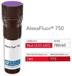
NB008214

CEACAM5/CD66e Antibody (C66/1291), DyLight 594, Novus Biologicals™

Manufacturer: Novus Biologicals

Select a Size

Pack Size SKU Availability Price
Each of 1 NB008214-Each-of-1 In Stock ₹ 53,688.90

NB008214 - Each of 1

₹ 53,688.90

In Stock

Quantity

1

Base Price: ₹ 53,688.90

GST (18%): ₹ 9,664.002

Total Price: ₹ 63,352.902

Antigen

CEACAM5/CD66e

Classification

Monoclonal

Conjugate

DyLight 594

Formulation

50mM Sodium Borate with 0.05% Sodium Azide

Gene Symbols

CEACAM5

Immunogen

Recombinant full-length human CEACAM5/CD66e protein (Uniprot: P06731)

Quantity

0.1 mL

Research Discipline

Apoptosis, Cancer, Cellular Markers, Immunology

Test Specificity

This antibody recognizes proteins of 80-200kDa, identified as different members of CEA family. CEA is synthesized during development in the fetal gut and is re-expressed in increased amounts in intestinal carcinomas and several other tumors. This monoclonal antibody does not react with nonspecific cross-reacting antigen (NCA) and with human polymorphonuclear leucocytes. It shows no reaction with a variety of normal tissues and is suitable for staining of formalin/paraffin tissues. CEA is not found in benign glands, stroma, or malignant prostatic cells. Antibody to CEA is useful in detecting early foci of gastric carcinoma and in distinguishing pulmonary adenocarcinomas (60-70% are CEA+) from pleural mesotheliomas (rarely or weakly CEA+). Anti-CEA positivity is seen in adenocarcinomas from the lung, colon, stomach, esophagus, pancreas, gallbladder, urachus, salivary gland, ovary, and endocervix.

Content And Storage

Store at 4°C in the dark.

Applications

Immunocytochemistry, Immunofluorescence, Immunohistochemistry (Paraffin)

Clone

C66/1291

Dilution

Immunocytochemistry/Immunofluorescence, Immunohistochemistry-Paraffin

Gene Alias

Carcinoembryonic antigen, carcinoembryonic antigen-related cell adhesion molecule 5, CD66e antigen, CEACD66e, DKFZp781M2392, Meconium antigen 100

Host Species

Mouse

Purification Method

Protein A or G purified

Regulatory Status

RUO

Primary or Secondary

Primary

Target Species

Human

Isotype

IgG1

Related Products

Img

Novus Biologicals

NB008190

--

Img

Novus Biologicals

NB008412

--

Img

Novus Biologicals

NB008057

--

Img

Novus Biologicals

NB008059

--

Img

Novus Biologicals

NB008310

--

Img

Novus Biologicals

NB005750

--

Img

Novus Biologicals

NB008053

--

Img

Novus Biologicals

NB005719

--

Description

  • CEACAM5/CD66e Monoclonal specifically detects CEACAM5/CD66e in Human samples
  • It is validated for ELISA, Immunohistochemistry-Paraffin.

Compare Similar Items

Show Difference

Img

Novus Biologicals

NB008214

--


Antigen:
CEACAM5/CD66e

Classification:
Monoclonal

Conjugate:
DyLight 594

Formulation:
50mM Sodium Borate with 0.05% Sodium Azide

Gene Symbols:
CEACAM5

Immunogen:
Recombinant full-length human CEACAM5/CD66e protein (Uniprot: P06731)

Quantity:
0.1 mL

Research Discipline:
Apoptosis, Cancer, Cellular Markers, Immunology

Test Specificity:
This antibody recognizes proteins of 80-200kDa, identified as different members of CEA family. CEA is synthesized during development in the fetal gut and is re-expressed in increased amounts in intestinal carcinomas and several other tumors. This monoclonal antibody does not react with nonspecific cross-reacting antigen (NCA) and with human polymorphonuclear leucocytes. It shows no reaction with a variety of normal tissues and is suitable for staining of formalin/paraffin tissues. CEA is not found in benign glands, stroma, or malignant prostatic cells. Antibody to CEA is useful in detecting early foci of gastric carcinoma and in distinguishing pulmonary adenocarcinomas (60-70% are CEA+) from pleural mesotheliomas (rarely or weakly CEA+). Anti-CEA positivity is seen in adenocarcinomas from the lung, colon, stomach, esophagus, pancreas, gallbladder, urachus, salivary gland, ovary, and endocervix.

Content And Storage:
Store at 4°C in the dark.

Applications:
Immunocytochemistry, Immunofluorescence, Immunohistochemistry (Paraffin)

Clone:
C66/1291

Dilution:
Immunocytochemistry/Immunofluorescence, Immunohistochemistry-Paraffin

Gene Alias:
Carcinoembryonic antigen, carcinoembryonic antigen-related cell adhesion molecule 5, CD66e antigen, CEACD66e, DKFZp781M2392, Meconium antigen 100

Host Species:
Mouse

Purification Method:
Protein A or G purified

Regulatory Status:
RUO

Primary or Secondary:
Primary

Target Species:
Human

Isotype:
IgG1

Img

Novus Biologicals

NB008218

--


Antigen:
CD11c

Classification:
Monoclonal

Conjugate:
DyLight 594

Formulation:
50mM Sodium Borate with 0.05% Sodium Azide

Gene Symbols:
ITGAX

Immunogen:
Recombinant fragment (aa 637-827) of human CD11c protein (exact sequence is proprietary) (Uniprot: P20702)

Quantity:
0.1 mL

Research Discipline:
Dendritic Cell Markers, Signal Transduction

Test Specificity:
Recognizes a protein of 145kDa, identified as CD11c. CD11c (ITGAX), a member of the leukointegrin family, shares the same beta subunit with other members of the leukocyte adhesion molecule family, which includes CD11a (LFA-1), CD11b (MAC-1) and CD11d (ITGAD), but has a unique alpha chain. CD11c has been shown to play a role in phagocytosis, cell migration, and cytokine production by monocytes/macrophages as well as induction of T-cell proliferation by Langerhans cells. CD11c is expressed prominently on the plasma membranes of monocytes, tissue macrophages, NK cells, and most dendritic cells (DCs). A lower level of expression is also observed on neutrophils as a result of its high level of expression on most DCs. An antibody to CD11c may aid in identification of lesions with histiocytic origin. It may also been used as a marker for hairy cell leukemia in paraffin-embedded tissues.

Content And Storage:
Store at 4°C in the dark.

Applications:
Flow Cytometry, Immunohistochemistry, Immunocytochemistry, Immunofluorescence, Immunohistochemistry (Paraffin)

Clone:
ITGAX/1243

Dilution:
Flow Cytometry, Immunohistochemistry, Immunocytochemistry/Immunofluorescence, Immunohistochemistry-Paraffin

Gene Alias:
95, 95 alpha chain, 95, alpha subunit, CD11 antigen-like family member C, CD11c, CD11c antigen, CD11Cleu M5, alpha subunit, integrin alpha-X, integrin, alpha X (antigen CD11C (p150), alpha polypeptide), integrin, alpha X (complement component 3 receptor 4 subunit), Leu M5, Leukocyte adhesion glycoprotein p150, Leukocyte adhesion receptor p150, leukocyte surface antigen p150, myeloid membrane antigen, alpha subunit, p150 95 integrin alpha chain, SLEB6

Host Species:
Mouse

Purification Method:
Protein A or G purified

Regulatory Status:
RUO

Primary or Secondary:
Primary

Target Species:
Human

Isotype:
IgG2b κ

Img

Novus Biologicals

NB008219

--


Antigen:
CD11c

Classification:
Monoclonal

Conjugate:
DyLight 488

Formulation:
50mM Sodium Borate with 0.05% Sodium Azide

Gene Symbols:
ITGAX

Immunogen:
Recombinant fragment (aa 637-827) of human CD11c protein (exact sequence is proprietary) (Uniprot: P20702)

Quantity:
0.1 mL

Research Discipline:
Dendritic Cell Markers, Signal Transduction

Test Specificity:
Recognizes a protein of 145kDa, identified as CD11c. CD11c (ITGAX), a member of the leukointegrin family, shares the same beta subunit with other members of the leukocyte adhesion molecule family, which includes CD11a (LFA-1), CD11b (MAC-1) and CD11d (ITGAD), but has a unique alpha chain. CD11c has been shown to play a role in phagocytosis, cell migration, and cytokine production by monocytes/macrophages as well as induction of T-cell proliferation by Langerhans cells. CD11c is expressed prominently on the plasma membranes of monocytes, tissue macrophages, NK cells, and most dendritic cells (DCs). A lower level of expression is also observed on neutrophils as a result of its high level of expression on most DCs. An antibody to CD11c may aid in identification of lesions with histiocytic origin. It may also been used as a marker for hairy cell leukemia in paraffin-embedded tissues.

Content And Storage:
Store at 4°C in the dark.

Applications:
Flow Cytometry, Immunohistochemistry, Immunocytochemistry, Immunofluorescence, Immunohistochemistry (Paraffin)

Clone:
ITGAX/1243

Dilution:
Flow Cytometry, Immunohistochemistry, Immunocytochemistry/Immunofluorescence, Immunohistochemistry-Paraffin

Gene Alias:
95, 95 alpha chain, 95, alpha subunit, CD11 antigen-like family member C, CD11c, CD11c antigen, CD11Cleu M5, alpha subunit, integrin alpha-X, integrin, alpha X (antigen CD11C (p150), alpha polypeptide), integrin, alpha X (complement component 3 receptor 4 subunit), Leu M5, Leukocyte adhesion glycoprotein p150, Leukocyte adhesion receptor p150, leukocyte surface antigen p150, myeloid membrane antigen, alpha subunit, p150 95 integrin alpha chain, SLEB6

Host Species:
Mouse

Purification Method:
Protein A or G purified

Regulatory Status:
RUO

Primary or Secondary:
Primary

Target Species:
Human

Isotype:
IgG2b κ

Img

Novus Biologicals

NB008220

--


Antigen:
PTH

Classification:
Monoclonal

Conjugate:
DyLight 488

Formulation:
50mM Sodium Borate with 0.05% Sodium Azide

Gene Symbols:
PTH

Immunogen:
A synthetic peptide around amino acids 1-34 of human mature-PTH-polypeptide (exact sequence is proprietary) (Uniprot: P01270)

Quantity:
0.1 mL

Research Discipline:
Apoptosis, Biologically Active Proteins, Cancer

Test Specificity:
Epitope of this monoclonal antibody maps in the N-terminus of PTH, a hormone produced by the parathyroid gland that regulates the concentration of calcium and phosphorus in extracellular fluid. This hormone elevates blood Ca2+ levels by dissolving the salts in bone and preventing their renal excretion. It is produced in the parathyroid gland as an 84 amino acid single chain polypeptide. It can also be secreted as N-terminal truncated fragments or C-terminal fragments after intracellular degradation, as in case of hypercalcemia. Defects in this gene are a cause of familial isolated hypoparathyroidism (FIH); also called autosomal dominant hypoparathyroidism or autosomal dominant hypocalcemia. FIH is characterized by hypocalcemia and hyperphosphatemia due to inadequate secretion of parathyroid hormone. Symptoms are seizures, tetany and cramps. FIH exist both as autosomal dominant and recessive forms of hypoparathyroidism.

Content And Storage:
Store at 4°C in the dark.

Applications:
Flow Cytometry, Immunohistochemistry, Immunocytochemistry, Immunofluorescence, Immunohistochemistry (Paraffin)

Clone:
PTH/911

Dilution:
Flow Cytometry, Immunohistochemistry, Immunocytochemistry/Immunofluorescence, Immunohistochemistry-Paraffin

Gene Alias:
Parathormone, Parathyrin, parathyroid hormone, parathyroid hormone 1, PTH1

Host Species:
Mouse

Purification Method:
Protein A or G purified

Regulatory Status:
RUO

Primary or Secondary:
Primary

Target Species:
Human

Isotype:
IgG2b κ