Histone H1 Antibody (21NC85), Novus Biologicals™
Manufacturer: Novus Biologicals
Select a Size
| Pack Size | SKU | Availability | Price |
|---|---|---|---|
| Each of 1 | NB10065220-Each-of-1 | In Stock | ₹ 43,387.50 |
NB10065220 - Each of 1
In Stock
Quantity
1
Base Price: ₹ 43,387.50
GST (18%): ₹ 7,809.75
Total Price: ₹ 51,197.25
Antigen
Histone H1
Classification
Monoclonal
Conjugate
Unconjugated
Gene Alias
H1 histone family, member 1, H1.1, H1A, H1F1HIST1, histone 1, H1a, histone cluster 1, H1a, histone H1.1, MGC126642, MGC138345
Host Species
Mouse
Purification Method
Affinity Purified
Regulatory Status
RUO
Gene ID (Entrez)
3024
Target Species
Human, Rat, Porcine
Isotype
IgG2a
Applications
Western Blot, ELISA, Immunohistochemistry, Immunocytochemistry, Immunofluorescence, Immunohistochemistry (Paraffin)
Clone
21NC85
Dilution
Western Blot 1:100-1:2000, ELISA 1:100-1:2000, Immunohistochemistry 1:10-1:500, Immunocytochemistry/Immunofluorescence 1:20-1:30, Immunohistochemistry-Paraffin 1:500, Immunohistochemistry-Frozen 1:20-1:50
Gene Symbols
H1-1
Immunogen
The exact immunogen is proprietary, but is composed of lysed nuclei of myeloid leukemia biopsy cells.
Quantity
0.05 mL
Primary or Secondary
Primary
Test Specificity
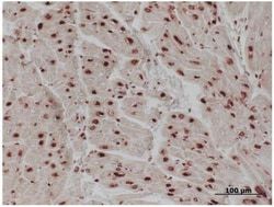
This antibody recognises an antigen found in the nucleus. Antibody also preferentially reacts with histone H1/ DNA nucleosome as shown by ELISA and western blotting. In WB using non-reduced samples it recognises primarily a 30-32kDa histone H1 band, but other bands are present as well. This antibody stains nuclei of all human cell types and also stains chromosomes diffusely in metaphase cells. Intense diffuse staining is obtained in interphase cells. The epitope has not been mapped.
Content And Storage
Store at -20C. Avoid freeze-thaw cycles.
Related Products


Description
- Description Histone H1 Monoclonal specifically detects Histone H1 in Human, Rat, Porcine samples
- It is validated for Western Blot, ELISA, Immunohistochemistry, Immunocytochemistry/Immunofluorescence, Immunohistochemistry-Paraffin, Immunohistochemistry-Frozen.
Compare Similar Items
Show Difference
Antigen: Histone H1
Classification: Monoclonal
Conjugate: Unconjugated
Gene Alias: H1 histone family, member 1, H1.1, H1A, H1F1HIST1, histone 1, H1a, histone cluster 1, H1a, histone H1.1, MGC126642, MGC138345
Host Species: Mouse
Purification Method: Affinity Purified
Regulatory Status: RUO
Gene ID (Entrez): 3024
Target Species: Human, Rat, Porcine
Isotype: IgG2a
Applications: Western Blot, ELISA, Immunohistochemistry, Immunocytochemistry, Immunofluorescence, Immunohistochemistry (Paraffin)
Clone: 21NC85
Dilution: Western Blot 1:100-1:2000, ELISA 1:100-1:2000, Immunohistochemistry 1:10-1:500, Immunocytochemistry/Immunofluorescence 1:20-1:30, Immunohistochemistry-Paraffin 1:500, Immunohistochemistry-Frozen 1:20-1:50
Gene Symbols: H1-1
Immunogen: The exact immunogen is proprietary, but is composed of lysed nuclei of myeloid leukemia biopsy cells.
Quantity: 0.05 mL
Primary or Secondary: Primary
Test Specificity: This antibody recognises an antigen found in the nucleus. Antibody also preferentially reacts with histone H1/ DNA nucleosome as shown by ELISA and western blotting. In WB using non-reduced samples it recognises primarily a 30-32kDa histone H1 band, but other bands are present as well. This antibody stains nuclei of all human cell types and also stains chromosomes diffusely in metaphase cells. Intense diffuse staining is obtained in interphase cells. The epitope has not been mapped.
Content And Storage: Store at -20C. Avoid freeze-thaw cycles.
Antigen: Coagulation Factor III/Tissue Factor
Classification: Monoclonal
Conjugate: Unconjugated
Gene Alias: CD142, CD142 antigen, Coagulation factor III, coagulation factor III (thromboplastin, tissue factor), FLJ17960, TF, TFA, Thromboplastin, tissue factor
Host Species: Mouse
Purification Method: Protein G purified
Regulatory Status: RUO
Gene ID (Entrez): 2152
Target Species: Human, Primate
Isotype: IgG1
Applications: Western Blot, Flow Cytometry, Immunohistochemistry, Immunocytochemistry, Immunofluorescence, Immunohistochemistry (Frozen)
Clone: TF9-10H10
Dilution: Western Blot 1-10 ug/ml, Flow Cytometry 1:10-1:1000, Immunohistochemistry 1:10-1:500, Immunocytochemistry/Immunofluorescence 1:10-1:500, Immunohistochemistry-Frozen 1:10-1:500
Gene Symbols: F3
Immunogen: Denatured Tissue factor isolated from human brain by the Factor VII affinity method.
Quantity: 0.1 mg
Primary or Secondary: Primary
Test Specificity: CD142, also known as Tissue Factor, is the membrane receptor for coagulation factors VII and VIIa and is the cell surface initiator of coagulation. It is the major molecule of this type and is criticial for controlling hemostasis, thrombosis and inflammation. NB100-65221 is specific for an epitope within the extracellular domain, epitope locus I. It recognizes both the reduced and native non-reduced human and primate tissue factors. It does not inhibit coagulation or neutralize factor VII binding to CD142.
Content And Storage: Store at 4C short term. Aliquot and store at -20C long term. Avoid freeze-thaw cycles.
Antigen: CD2
Classification: Monoclonal
Conjugate: DyLight 488
Gene Alias: CD2 antigen, CD2 antigen (p50), sheep red blood cell receptor, CD2 molecule, Erythrocyte receptor, FLJ46032, LFA-2, LFA-3 receptor, lymphocyte-function antigen-2, Rosette receptor, SRBC, T11, T-cell surface antigen CD2, T-cell surface antigen T11/Leu-5
Host Species: Mouse
Purification Method: Protein G purified
Regulatory Status: RUO
Gene ID (Entrez): 914
Target Species: Rat
Isotype: IgG2a
Applications: Flow Cytometry, Immunocytochemistry, Immunofluorescence, Immunohistochemistry (Paraffin), Immunohistochemistry (Frozen)
Clone: OX-34
Dilution: Flow Cytometry, Immunocytochemistry/Immunofluorescence, Immunohistochemistry-Paraffin, Immunohistochemistry-Frozen
Gene Symbols: CD2
Immunogen: Activated rat T helper cells
Quantity: 0.25 mL
Primary or Secondary: Primary
Test Specificity: NB100-65228 recognizes the rat CD2 cell surface antigen, a 50-54kD glycoprotein expressed by thymocytes and mature T cells. * Whiteland, J.L. et al. (1995). Immunohistochemical Detection of T-Cell Subsets and other Leucocytes in Paraffin-embedded Rat and Mouse Tissues with Monoclonal Antibodies. J. Histochem. Cytochem. 43: 313-320.
Content And Storage: Store at 4C in the dark.