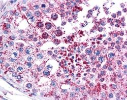
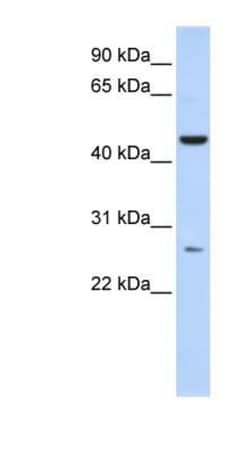
AKD1 Antibody, Novus Biologicals™
Manufacturer: Novus Biologicals
Select a Size
| Pack Size | SKU | Availability | Price |
|---|---|---|---|
| Each of 1 | NBP156395-Each-of-1 | In Stock | ₹ 43,387.50 |
NBP156395 - Each of 1
In Stock
Quantity
1
Base Price: ₹ 43,387.50
GST (18%): ₹ 7,809.75
Total Price: ₹ 51,197.25
Antigen
AKD1
Classification
Polyclonal
Conjugate
Unconjugated
Formulation
PBS, 2% Sucrose with 0.09% Sodium Azide
Gene Alias
adenylate kinase domain containing 1, adenylate kinase domain containing 2, adenylate kinase domain-containing protein 1, Adenylate kinase domain-containing protein 2, AKD2, C6orf199, C6orf224, chromosome 6 open reading frame 199, chromosome 6 open reading frame 224, dJ70A9.1, FLJ16163, FLJ25791, FLJ34784, FLJ42177, MGC126763, MGC138153, MGC177059, MGC180194, MGC184281, MGC26954, RP1-70A9.1
Host Species
Rabbit
Purification Method
Affinity purified
Regulatory Status
RUO
Primary or Secondary
Primary
Test Specificity
Expected identity based on immunogen sequence: Crab-eating macaque: 100%; Human: 100%;.
Target Species
Human, Rat, Bovine, Canine, Equine, Guinea Pig, Rabbit
Isotype
IgG
Applications
Western Blot
Concentration
0.5 mg/ml
Dilution
Western Blot 1.0 ug/ml
Gene Accession No.
Q5TCS8
Gene Symbols
AK9
Immunogen
Synthetic peptides corresponding to C6ORF199 The peptide sequence was selected from the middle region of C6ORF199. Peptide sequence IINIKCPDYDLCQRISGQRQHNNTGYIYSRDQWDPEVIENHRKKKKEAQK.
Quantity
100 μL
Research Discipline
Protein Kinase
Gene ID (Entrez)
221264
Reconstitution
Centrifuge the vial of lyoph antibody at 12,000 x g for 20 seconds. Add 50μL of distilled water. Vortex followed by centrifuge again to pellet the solution.Final concentration is 1mg/mL in PBS buffer.
Content And Storage
Store at 4°C short term. Aliquot and store at -20°C long term. Avoid freeze-thaw cycles.
Description
- AKD1 Polyclonal specifically detects AKD1 in Human samples
- It is validated for Western Blot.
Compare Similar Items
Show Difference
Antigen: AKD1
Classification: Polyclonal
Conjugate: Unconjugated
Formulation: PBS, 2% Sucrose with 0.09% Sodium Azide
Gene Alias: adenylate kinase domain containing 1, adenylate kinase domain containing 2, adenylate kinase domain-containing protein 1, Adenylate kinase domain-containing protein 2, AKD2, C6orf199, C6orf224, chromosome 6 open reading frame 199, chromosome 6 open reading frame 224, dJ70A9.1, FLJ16163, FLJ25791, FLJ34784, FLJ42177, MGC126763, MGC138153, MGC177059, MGC180194, MGC184281, MGC26954, RP1-70A9.1
Host Species: Rabbit
Purification Method: Affinity purified
Regulatory Status: RUO
Primary or Secondary: Primary
Test Specificity: Expected identity based on immunogen sequence: Crab-eating macaque: 100%; Human: 100%;.
Target Species: Human, Rat, Bovine, Canine, Equine, Guinea Pig, Rabbit
Isotype: IgG
Applications: Western Blot
Concentration: 0.5 mg/ml
Dilution: Western Blot 1.0 ug/ml
Gene Accession No.: Q5TCS8
Gene Symbols: AK9
Immunogen: Synthetic peptides corresponding to C6ORF199 The peptide sequence was selected from the middle region of C6ORF199. Peptide sequence IINIKCPDYDLCQRISGQRQHNNTGYIYSRDQWDPEVIENHRKKKKEAQK.
Quantity: 100 μL
Research Discipline: Protein Kinase
Gene ID (Entrez): 221264
Reconstitution: Centrifuge the vial of lyoph antibody at 12,000 x g for 20 seconds. Add 50μL of distilled water. Vortex followed by centrifuge again to pellet the solution.Final concentration is 1mg/mL in PBS buffer.
Content And Storage: Store at 4°C short term. Aliquot and store at -20°C long term. Avoid freeze-thaw cycles.
Antigen: XPG
Classification: Polyclonal
Conjugate: Unconjugated
Formulation: PBS, 2% Sucrose with 0.09% Sodium Azide
Gene Alias: COFS3, DNA excision repair protein ERCC-5, DNA repair protein complementing XP-G cells, EC 3.1, ERCM2, excision repair cross-complementing rodent repair deficiency, complementationgroup 5, excision repair protein, xeroderma pigmentosum complementation group G protein, Xeroderma pigmentosum group G-complementing protein, xeroderma pigmentosum, complementation group G, XPGC, XPG-complementing protein, XPGUVDR
Host Species: Rabbit
Purification Method: Affinity purified
Regulatory Status: RUO
Primary or Secondary: Primary
Test Specificity: Expected identity based on immunogen sequence: Human: 100%; Mouse: 100%; Rabbit: 100%; Rat: 100%; Guinea pig: 92%; Pig: 92%; Xenopus: 85%; Bovine: 85%; Canine: 85%; Equine: 85%; Chicken: 78%.
Target Species: Human, Mouse, Rat, Bovine, Canine, Equine, Guinea Pig, Rabbit
Isotype: IgG
Applications: Western Blot
Concentration: 0.5 mg/ml
Dilution: Western Blot 1.0 ug/ml
Gene Accession No.: P28715
Gene Symbols: ERCC5
Immunogen: Synthetic peptide corresponding to the N terminal of ERCC5(excision repair cross-complementing rodent repair deficiency, complementation group 5 (xeroderma pigmentosum, complementation group G (Cockayne syndrome))) Peptide sequence NPQAIDIESEDFSSLPPEVKHEILTDMKEFTKRRRTLFEAMPEESDDFSQ The peptide sequence for this immunogen was taken from within the described region.
Quantity: 100 μL
Research Discipline: Cancer, DNA Repair, Nucleotide Excision Repair
Gene ID (Entrez): 2073
Reconstitution: Centrifuge the vial of lyoph antibody at 12,000 x g for 20 seconds. Add 50μL of distilled water. Vortex followed by centrifuge again to pellet the solution.Final concentration is 1mg/mL in PBS buffer.
Content And Storage: Store at 4°C short term. Aliquot and store at -20°C long term. Avoid freeze-thaw cycles.
Antigen: ASPA
Classification: Polyclonal
Conjugate: Unconjugated
Formulation: PBS, 2% Sucrose with 0.09% Sodium Azide
Gene Alias: ACY-2, ACY2aminoacylase 2, Aminoacylase-2, ASPaminoacylase-2, aspartoacylase, aspartoacylase (aminoacylase 2, Canavan disease), EC 3.5.1.15
Host Species: Rabbit
Purification Method: Affinity purified
Regulatory Status: RUO
Primary or Secondary: Primary
Test Specificity: Expected identity based on immunogen sequence: Bovine: 100%; Canine: 100%; Guinea pig: 100%; Equine: 100%; Human: 100%; Mouse: 100%; Rat: 100%; Chicken: 92%.
Target Species: Human, Mouse, Rat, Bovine, Canine, Equine, Guinea Pig, Rabbit, Zebrafish
Isotype: IgG
Applications: Western Blot
Concentration: 0.5 mg/ml
Dilution: Western Blot 1.0 ug/ml
Gene Accession No.: P45381
Gene Symbols: ASPA
Immunogen: Synthetic peptides corresponding to ASPA(aspartoacylase (Canavan disease)) The peptide sequence was selected from the N terminal of ASPA. Peptide sequence RIFDLENLGKKMSEDLPYEVRRAQEINHLFGPKDSEDSYDIIFDLHNTTS.
Quantity: 100 μL
Research Discipline: Neuroscience
Gene ID (Entrez): 443
Reconstitution: Centrifuge the vial of lyoph antibody at 12,000 x g for 20 seconds. Add 50μL of distilled water. Vortex followed by centrifuge again to pellet the solution.Final concentration is 1mg/mL in PBS buffer.
Content And Storage: Store at 4°C short term. Aliquot and store at -20°C long term. Avoid freeze-thaw cycles.