Cytokeratin 19 Antibody (E6), Novus Biologicals™
Manufacturer: Fischer Scientific
Select a Size
| Pack Size | SKU | Availability | Price |
|---|---|---|---|
| Each of 1 | NBP229804-Each-of-1 | In Stock | ₹ 52,065.00 |
NBP229804 - Each of 1
In Stock
Quantity
1
Base Price: ₹ 52,065.00
GST (18%): ₹ 9,371.70
Total Price: ₹ 61,436.70
Antigen
Cytokeratin 19
Classification
Monoclonal
Conjugate
Unconjugated
Formulation
Ascites with No Preservative
Gene Alias
CK19, CK-19, cytokeratin 19, Cytokeratin-19,40-kDa keratin intermediate filament, K19cytokeratin-19, K1CS, keratin 19, keratin, type I cytoskeletal 19, keratin, type I, 40-kd, keratin-19, MGC15366
Host Species
Mouse
Purification Method
Unpurified
Regulatory Status
RUO
Primary or Secondary
Primary
Test Specificity
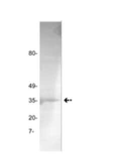
Rat keratin 19 (40kD), analogous to human keratin 19.
Content And Storage
Aliquot and store at -20C or -80C. Avoid freeze-thaw cycles.
Isotype
IgG1
Applications
Western Blot, Immunohistochemistry, Immunohistochemistry (Frozen)
Clone
E6
Dilution
Western Blot 1:100-1:2000, Immunohistochemistry 1:10-1:500, Immunohistochemistry-Frozen
Gene Accession No.
P08727, Q63279
Gene Symbols
KRT19
Immunogen
Keratin from rat colon.
Quantity
0.1 mL
Research Discipline
Cancer, Cell Biology, Cellular Markers, Cytoskeleton Markers, Neuroscience, Stem Cell Markers
Gene ID (Entrez)
3880
Target Species
Rat, Fish, Rabbit, Human (Negative), Mouse (Negative)
Form
Ascites
Related Products


Description
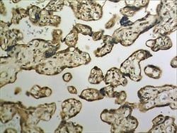
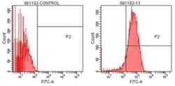
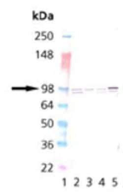
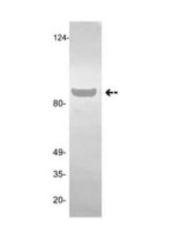
- Cytokeratin 19 Monoclonal specifically detects Cytokeratin 19 in Rat, Fish, Rabbit, Human (Negative), Mouse (Negative) samples
- It is validated for Western Blot, Immunohistochemistry, Immunocytochemistry/Immunofluorescence, Immunohistochemistry-Frozen.
Compare Similar Items
Show Difference
Antigen: Cytokeratin 19
Classification: Monoclonal
Conjugate: Unconjugated
Formulation: Ascites with No Preservative
Gene Alias: CK19, CK-19, cytokeratin 19, Cytokeratin-19,40-kDa keratin intermediate filament, K19cytokeratin-19, K1CS, keratin 19, keratin, type I cytoskeletal 19, keratin, type I, 40-kd, keratin-19, MGC15366
Host Species: Mouse
Purification Method: Unpurified
Regulatory Status: RUO
Primary or Secondary: Primary
Test Specificity: Rat keratin 19 (40kD), analogous to human keratin 19.
Content And Storage: Aliquot and store at -20C or -80C. Avoid freeze-thaw cycles.
Isotype: IgG1
Applications: Western Blot, Immunohistochemistry, Immunohistochemistry (Frozen)
Clone: E6
Dilution: Western Blot 1:100-1:2000, Immunohistochemistry 1:10-1:500, Immunohistochemistry-Frozen
Gene Accession No.: P08727, Q63279
Gene Symbols: KRT19
Immunogen: Keratin from rat colon.
Quantity: 0.1 mL
Research Discipline: Cancer, Cell Biology, Cellular Markers, Cytoskeleton Markers, Neuroscience, Stem Cell Markers
Gene ID (Entrez): 3880
Target Species: Rat, Fish, Rabbit, Human (Negative), Mouse (Negative)
Form: Ascites
Antigen: Angiostatin
Classification: Monoclonal
Conjugate: Unconjugated
Formulation: No buffer with No Preservative
Gene Alias: __
Host Species: Mouse
Purification Method: Affinity Purified
Regulatory Status: RUO
Primary or Secondary: Primary
Test Specificity: Plasminogen (kringles 1-3).
Content And Storage: Aliquot and store at -20C or -80C. Avoid freeze-thaw cycles.
Isotype: IgM
Applications: Western Blot, ELISA
Clone: GMA-013
Dilution: Western Blot 1:100-1:2000, ELISA 1:100-1:2000
Gene Accession No.: P00747
Gene Symbols: PLG
Immunogen: Purified human plasminogen.
Quantity: 0.1 mg
Research Discipline: __
Gene ID (Entrez): 5340
Target Species: Human
Form: __
Antigen: Coagulation Factor II/Thrombin
Classification: Monoclonal
Conjugate: Unconjugated
Formulation: No buffer with No Preservative
Gene Alias: Coagulation Factor II, coagulation factor II (thrombin) receptor-like 2, Coagulation factor II receptor-like 2, Coagulation factor II receptor-like 2 (protease-actovated receptor 3), PAR-3, PAR3proteinase-activated receptor 3, protease-activated receptor 3, proteinase-activated receptor-3, PT, serine protease, Thrombin receptor-like 2
Host Species: Mouse
Purification Method: Affinity Purified
Regulatory Status: RUO
Primary or Secondary: Primary
Test Specificity: __
Content And Storage: Aliquot and store at -20C or -80C. Avoid freeze-thaw cycles.
Isotype: IgG1
Applications: Western Blot
Clone: GMA-020
Dilution: Western Blot 1:100-1:2000
Gene Accession No.: P00734
Gene Symbols: F2
Immunogen: purified human thrombin.
Quantity: 0.1 mg
Research Discipline: Apoptosis, Breast Cancer, Cancer, GPCR, Signal Transduction
Gene ID (Entrez): 2147
Target Species: Human
Form: __