CD34 Mouse, Clone: SPM123, Novus Biologicals™
Manufacturer: Fischer Scientific
Select a Size
| Pack Size | SKU | Availability | Price |
|---|---|---|---|
| Each of 1 | NBP232933A-Each-of-1 | In Stock | ₹ 47,704.00 |
NBP232933A - Each of 1
In Stock
Quantity
1
Base Price: ₹ 47,704.00
GST (18%): ₹ 8,586.72
Total Price: ₹ 56,290.72
Antigen
CD34
Classification
Monoclonal
Conjugate
Unconjugated
Formulation
PBS with 0.05% BSA. with 0.05% Sodium Azide
Gene Alias
CD34 antigenhematopoietic progenitor cell antigen CD34, CD34 molecule
Host Species
Mouse
Purification Method
Protein A purified
Regulatory Status
RUO
Primary or Secondary
Primary
Test Specificity
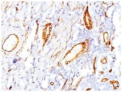
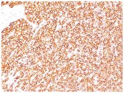
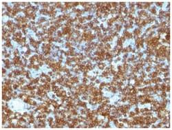
This MAb recognizes a single chain, transmembrane, heavily glycosylated protein of 90-120kDa, which is identified as CD34. On the basis of differential sensitivity to degradation by specific enzymes, epitopes of monoclonal antibodies to CD34 are classified into three main categories, class I, class II and class III. It is a class II antibody whose epitope is resistant to neuraminidase but sensitive to glycoprotease and chymopapain. CD34 expression is a hallmark for identifying pluripotent hematopoietic stem or progenitor cells. Its expression is gradually lost as lineage committed progenitors differentiate. CD34 is a marker of choice for staining blasts in acute myeloid leukemia. In addition, CD34 is expressed by soft tissue tumors, such as solitary fibrous tumor and gastrointestinal stromal tumor. Its expression is also found in vascular endothelium. Additionally, it appears that proliferating endothelial cells express this molecule more than the non-proliferating endothelial cells. A
Content And Storage
Store at 4C.
Isotype
IgG1 κ
Applications
Western Blot, Flow Cytometry, Immunocytochemistry, Immunofluorescence, Immunoprecipitation, Immunohistochemistry (Paraffin)
Clone
SPM123
Dilution
Western Blot 0.25-0.5ug/ml, Flow Cytometry 0.5-1ug/million cells, Immunocytochemistry/Immunofluorescence 0.5-1ug/ml, Immunoprecipitation 0.5-1ug/500ug protein lysate, Immunohistochemistry-Paraffin 0.5-1.0ug/ml, Immunohistochemistry-Frozen 0.5-1.0ug/ml
Gene Accession No.
P28906
Gene Symbols
CD34
Immunogen
Detergent solubilized vesicular suspension prepared from human term placenta
Quantity
0.1 mg
Research Discipline
Adaptive Immunity, Breast Cancer, Cancer, Cell Biology, Cellular Markers, Endothelial Cell Markers, Hematopoietic Stem Cell Markers, Hypoxia, Immunology, Innate Immunity, Mast Cell Markers, Mesenchymal Stem Cell Markers, Myeloid Cell Markers, Neuronal Cell Markers, Neuroscience, Stem Cell Markers, Tumor Suppressors
Gene ID (Entrez)
947
Target Species
Human, Primate, Bovine (Negative), Canine (Negative), Rat (Negative), Sheep (Negative)
Form
Purified
Related Products

Description
- CD34 Monoclonal specifically detects CD34 in Human, Cynomolgus Monkey, Rhesus Macaque, Bovine (Negative), Canine (Negative), Rat (Negative), Sheep (Negative) samples
- It is validated for Flow Cytometry, Immunohistochemistry, Immunocytochemistry/Immunofluorescence, Immunohistochemistry-Paraffin.
Compare Similar Items
Show Difference
Antigen: CD34
Classification: Monoclonal
Conjugate: Unconjugated
Formulation: PBS with 0.05% BSA. with 0.05% Sodium Azide
Gene Alias: CD34 antigenhematopoietic progenitor cell antigen CD34, CD34 molecule
Host Species: Mouse
Purification Method: Protein A purified
Regulatory Status: RUO
Primary or Secondary: Primary
Test Specificity: This MAb recognizes a single chain, transmembrane, heavily glycosylated protein of 90-120kDa, which is identified as CD34. On the basis of differential sensitivity to degradation by specific enzymes, epitopes of monoclonal antibodies to CD34 are classified into three main categories, class I, class II and class III. It is a class II antibody whose epitope is resistant to neuraminidase but sensitive to glycoprotease and chymopapain. CD34 expression is a hallmark for identifying pluripotent hematopoietic stem or progenitor cells. Its expression is gradually lost as lineage committed progenitors differentiate. CD34 is a marker of choice for staining blasts in acute myeloid leukemia. In addition, CD34 is expressed by soft tissue tumors, such as solitary fibrous tumor and gastrointestinal stromal tumor. Its expression is also found in vascular endothelium. Additionally, it appears that proliferating endothelial cells express this molecule more than the non-proliferating endothelial cells. A
Content And Storage: Store at 4C.
Isotype: IgG1 κ
Applications: Western Blot, Flow Cytometry, Immunocytochemistry, Immunofluorescence, Immunoprecipitation, Immunohistochemistry (Paraffin)
Clone: SPM123
Dilution: Western Blot 0.25-0.5ug/ml, Flow Cytometry 0.5-1ug/million cells, Immunocytochemistry/Immunofluorescence 0.5-1ug/ml, Immunoprecipitation 0.5-1ug/500ug protein lysate, Immunohistochemistry-Paraffin 0.5-1.0ug/ml, Immunohistochemistry-Frozen 0.5-1.0ug/ml
Gene Accession No.: P28906
Gene Symbols: CD34
Immunogen: Detergent solubilized vesicular suspension prepared from human term placenta
Quantity: 0.1 mg
Research Discipline: Adaptive Immunity, Breast Cancer, Cancer, Cell Biology, Cellular Markers, Endothelial Cell Markers, Hematopoietic Stem Cell Markers, Hypoxia, Immunology, Innate Immunity, Mast Cell Markers, Mesenchymal Stem Cell Markers, Myeloid Cell Markers, Neuronal Cell Markers, Neuroscience, Stem Cell Markers, Tumor Suppressors
Gene ID (Entrez): 947
Target Species: Human, Primate, Bovine (Negative), Canine (Negative), Rat (Negative), Sheep (Negative)
Form: Purified
Antigen: CD34
Classification: Monoclonal
Conjugate: Unconjugated
Formulation: PBS with 0.05% BSA. with 0.05% Sodium Azide
Gene Alias: CD34 antigenhematopoietic progenitor cell antigen CD34, CD34 molecule
Host Species: Mouse
Purification Method: Protein A purified
Regulatory Status: RUO
Primary or Secondary: Primary
Test Specificity: This MAb recognizes a single chain, transmembrane, heavily glycosylated protein of 90-120kDa, which is identified as CD34. On the basis of differential sensitivity to degradation by specific enzymes, epitopes of monoclonal antibodies to CD34 are classified into three main categories, class I, class II and class III. It is a class II antibody whose epitope is resistant to neuraminidase but sensitive to glycoprotease and chymopapain. CD34 expression is a hallmark for identifying pluripotent hematopoietic stem or progenitor cells. Its expression is gradually lost as lineage committed progenitors differentiate. CD34 is a marker of choice for staining blasts in acute myeloid leukemia. In addition, CD34 is expressed by soft tissue tumors, such as solitary fibrous tumor and gastrointestinal stromal tumor. Its expression is also found in vascular endothelium. Additionally, it appears that proliferating endothelial cells express this molecule more than the non-proliferating endothelial cells. A
Content And Storage: Store at 4C.
Isotype: IgG1 κ
Applications: Western Blot, Flow Cytometry, Immunocytochemistry, Immunofluorescence, Immunoprecipitation, Immunohistochemistry (Paraffin)
Clone: SPM123
Dilution: Western Blot 0.25-0.5ug/ml, Flow Cytometry 0.5-1ug/million cells, Immunocytochemistry/Immunofluorescence 0.5-1ug/ml, Immunoprecipitation 0.5-1ug/500ug protein lysate, Immunohistochemistry-Paraffin 0.5-1.0ug/ml, Immunohistochemistry-Frozen 0.5-1.0ug/ml
Gene Accession No.: P28906
Gene Symbols: CD34
Immunogen: Detergent solubilized vesicular suspension prepared from human term placenta
Quantity: 0.2 mg
Research Discipline: Adaptive Immunity, Breast Cancer, Cancer, Cell Biology, Cellular Markers, Endothelial Cell Markers, Hematopoietic Stem Cell Markers, Hypoxia, Immunology, Innate Immunity, Mast Cell Markers, Mesenchymal Stem Cell Markers, Myeloid Cell Markers, Neuronal Cell Markers, Neuroscience, Stem Cell Markers, Tumor Suppressors
Gene ID (Entrez): 947
Target Species: Human, Primate, Bovine (Negative), Canine (Negative), Rat (Negative), Sheep (Negative)
Form: Purified
Antigen: CD34
Classification: Monoclonal
Conjugate: Unconjugated
Formulation: PBS with 0.05% BSA. with 0.05% Sodium Azide
Gene Alias: CD34 antigenhematopoietic progenitor cell antigen CD34, CD34 molecule
Host Species: Mouse
Purification Method: Protein A purified
Regulatory Status: RUO
Primary or Secondary: Primary
Test Specificity: This MAb recognizes a single chain, transmembrane, heavily glycosylated protein of 90-120kDa, which is identified as CD34. On the basis of differential sensitivity to degradation by specific enzymes, epitopes of monoclonal antibodies to CD34 are classified into three main categories, class I, class II and class III. It is a class II antibody whose epitope is resistant to neuraminidase but sensitive to glycoprotease and chymopapain. CD34 expression is a hallmark for identifying pluripotent hematopoietic stem or progenitor cells. Its expression is gradually lost as lineage committed progenitors differentiate. CD34 is a marker of choice for staining blasts in acute myeloid leukemia. In addition, CD34 is expressed by soft tissue tumors, such as solitary fibrous tumor and gastrointestinal stromal tumor. Its expression is also found in vascular endothelium. Additionally, it appears that proliferating endothelial cells express this molecule more than the non-proliferating endothelial cells. A
Content And Storage: Store at 4C.
Isotype: IgG1 κ
Applications: Western Blot, Flow Cytometry, Immunocytochemistry, Immunofluorescence, Immunoprecipitation, Immunohistochemistry (Paraffin)
Clone: SPM123
Dilution: Western Blot 0.25-0.5ug/ml, Flow Cytometry 0.5-1ug/million cells, Immunocytochemistry/Immunofluorescence 0.5-1ug/ml, Immunoprecipitation 0.5-1ug/500ug protein lysate, Immunohistochemistry-Paraffin 0.5-1.0ug/ml, Immunohistochemistry-Frozen 0.5-1.0ug/ml
Gene Accession No.: P28906
Gene Symbols: CD34
Immunogen: Detergent solubilized vesicular suspension prepared from human term placenta
Quantity: 0.02 mg
Research Discipline: Adaptive Immunity, Breast Cancer, Cancer, Cell Biology, Cellular Markers, Endothelial Cell Markers, Hematopoietic Stem Cell Markers, Hypoxia, Immunology, Innate Immunity, Mast Cell Markers, Mesenchymal Stem Cell Markers, Myeloid Cell Markers, Neuronal Cell Markers, Neuroscience, Stem Cell Markers, Tumor Suppressors
Gene ID (Entrez): 947
Target Species: Human, Primate, Bovine (Negative), Canine (Negative), Rat (Negative), Sheep (Negative)
Form: Purified
