CD45RB Mouse, Clone: BRA-11 (same as BRA-11G), Novus Biologicals™
Mouse Monoclonal Antibody
Manufacturer: Fischer Scientific
The price for this product is unavailable. Please request a quote
Antigen
CD45RB
Dilution
Western Blot 0.5-1.0ug/ml, Flow Cytometry 0.5-1ug/million cells, Immunocytochemistry/Immunofluorescence 0.5-1ug/ml, Immunoprecipitation 0.5-1ug/500ug protein lysate, Immunohistochemistry-Paraffin 0.5-1.0ug/ml, Immunohistochemistry-Frozen 0.5-1.0ug/ml
Classification
Monoclonal
Form
Purified
Regulatory Status
RUO
Target Species
Human, Monkey (Negative), Rat (Negative)
Gene Accession No.
P08575
Gene ID (Entrez)
5788
Immunogen
Non-T, non-B CALLA positive ALL cell line REH (Leucocyte Workshop IV and V)
Primary or Secondary
Primary
Content And Storage
Store at 4C.
Clone
BRA-11 (same as BRA-11G)
Applications
Western Blot, Flow Cytometry, Immunocytochemistry, Immunofluorescence, Immunoprecipitation, Immunohistochemistry (Paraffin)
Conjugate
Unconjugated
Host Species
Mouse
Research Discipline
Adaptive Immunity, Cell Biology, Cellular Markers, Cytokine Research, Hematopoietic Stem Cell Markers, Immunology, Mesenchymal Stem Cell Markers, Microglia Markers, Neuronal Cell Markers, Neuroscience, Signal Transduction, Stem Cell Markers
Formulation
PBS with 0.05% BSA. with 0.05% Sodium Azide
Gene Alias
B220, CD45 antigen, CD45R, EC 3.1.3.48, L-CA, LY5, protein tyrosine phosphatase, receptor type, C, receptor-type tyrosine-protein phosphatase C, T200 glycoprotein, T200 leukocyte common antigen, T200receptor type, c polypeptide
Gene Symbols
PTPRC
Isotype
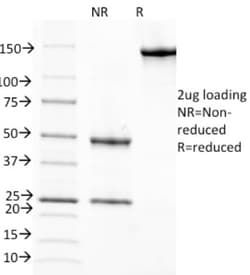
IgG1 κ
Purification Method
Protein A purified
Test Specificity
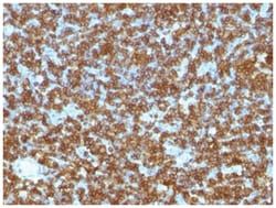
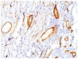
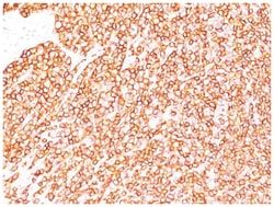
CD45R, also designated CD45 and PTPRC, has been identified as a transmembrane glycoprotein, broadly expressed among hematopoietic cells. Multiple isoforms of CD45R are distributed throughout the immune system according to cell type. These isoforms arise because of alternative splicing of exons 4, 5, and 6. The corresponding protein domains are characterized by the binding of monoclonal antibodies specific for CD45RA (exon 4), CD45RB (exon 5), CD45RC (exon 6) and CD45RO (exons 4 to 6 spliced out). The variation in these isoforms is localized to the extracellular domain of CD45R, while the intracellular domain is conserved. CD45RB is expressed on mature B-lymphocytes and the majority of lymphomas and leukemias of B-cell origin.
Related Products

Description
- CD45RB Monoclonal specifically detects CD45RB in Human, Monkey (Negative), Rat (Negative) samples
- It is validated for Western Blot, Flow Cytometry, Immunohistochemistry, Immunocytochemistry/Immunofluorescence, Immunohistochemistry-Paraffin.
Compare Similar Items
Show Difference
Antigen: CD45RB
Dilution: Western Blot 0.5-1.0ug/ml, Flow Cytometry 0.5-1ug/million cells, Immunocytochemistry/Immunofluorescence 0.5-1ug/ml, Immunoprecipitation 0.5-1ug/500ug protein lysate, Immunohistochemistry-Paraffin 0.5-1.0ug/ml, Immunohistochemistry-Frozen 0.5-1.0ug/ml
Classification: Monoclonal
Form: Purified
Regulatory Status: RUO
Target Species: Human, Monkey (Negative), Rat (Negative)
Gene Accession No.: P08575
Gene ID (Entrez): 5788
Immunogen: Non-T, non-B CALLA positive ALL cell line REH (Leucocyte Workshop IV and V)
Primary or Secondary: Primary
Content And Storage: Store at 4C.
Clone: BRA-11 (same as BRA-11G)
Applications: Western Blot, Flow Cytometry, Immunocytochemistry, Immunofluorescence, Immunoprecipitation, Immunohistochemistry (Paraffin)
Conjugate: Unconjugated
Host Species: Mouse
Research Discipline: Adaptive Immunity, Cell Biology, Cellular Markers, Cytokine Research, Hematopoietic Stem Cell Markers, Immunology, Mesenchymal Stem Cell Markers, Microglia Markers, Neuronal Cell Markers, Neuroscience, Signal Transduction, Stem Cell Markers
Formulation: PBS with 0.05% BSA. with 0.05% Sodium Azide
Gene Alias: B220, CD45 antigen, CD45R, EC 3.1.3.48, L-CA, LY5, protein tyrosine phosphatase, receptor type, C, receptor-type tyrosine-protein phosphatase C, T200 glycoprotein, T200 leukocyte common antigen, T200receptor type, c polypeptide
Gene Symbols: PTPRC
Isotype: IgG1 κ
Purification Method: Protein A purified
Test Specificity: CD45R, also designated CD45 and PTPRC, has been identified as a transmembrane glycoprotein, broadly expressed among hematopoietic cells. Multiple isoforms of CD45R are distributed throughout the immune system according to cell type. These isoforms arise because of alternative splicing of exons 4, 5, and 6. The corresponding protein domains are characterized by the binding of monoclonal antibodies specific for CD45RA (exon 4), CD45RB (exon 5), CD45RC (exon 6) and CD45RO (exons 4 to 6 spliced out). The variation in these isoforms is localized to the extracellular domain of CD45R, while the intracellular domain is conserved. CD45RB is expressed on mature B-lymphocytes and the majority of lymphomas and leukemias of B-cell origin.

Antigen: pan Actin
Dilution: Western Blot 0.5-1.0ug/ml, Simple Western 10 ug/ml, Flow Cytometry 0.5-1ug/million cells, Immunocytochemistry/Immunofluorescence 0.5-1ug/ml, Immunoprecipitation 0.5-1ug/500ug protein lysate, Immunohistochemistry-Paraffin 0.5-1ug/ml, Immunohistochemistry-Frozen 0.5-1ug/mlimmunohistochemistry-Paraffin 0.5-1ug/ml, SDS-Page
Classification: Monoclonal
Form: Purified
Regulatory Status: RUO
Target Species: Human, Mouse, Rat, Canine, Chicken, Feline, Rabbit
Gene Accession No.: P62736
Gene ID (Entrez): 58
Immunogen: SDS extract of human myocardium.
Primary or Secondary: Primary
Content And Storage: Store at 4C.
Clone: HHF35
Applications: Western Blot, Flow Cytometry, Immunocytochemistry, Immunofluorescence, Immunoprecipitation
Conjugate: Unconjugated
Host Species: Mouse
Research Discipline: __
Formulation: PBS with 0.05% BSA. with 0.05% Sodium Azide
Gene Alias: ACTA, actin, alpha 1, skeletal muscle, alpha skeletal muscle, alpha skeletal muscle actin, alpha-actin-1, ASMA, CFTD, CFTDM, MPFD, NEM1, NEM2, NEM3
Gene Symbols: ACTA1
Isotype: IgG1 κ
Purification Method: Protein A purified
Test Specificity: This antibody recognizes actin of skeletal, cardiac, and smooth muscle cells. It is not reactive with other mesenchymal cells except for myoepithelium. Actin can be resolved on the basis of its isoelectric points into three distinctive components: alpha, beta, and gamma in order of increasing isoelectric point. Anti-muscle specific actin recognizes alpha and gamma isotypes of all muscle groups. Non-muscle cells such as vascular endothelial cells and connective tissues are non-reactive. Also, neoplastic cells of non-muscle-derived tissue such as carcinomas, melanomas, and lymphomas are negative.It stains tumors of smooth muscle (leiomyomas and leiomyosarcomas) as well as skeletal muscle (rhabdomyomas and rhabdomyosarcomas).
Antigen: Retinol Binding Protein RBP
Dilution: Western Blot 0.5-1ug/ml, Immunocytochemistry/Immunofluorescence 1-2ug/ml, Immunoprecipitation 1-2ug/500ug protein, Immunohistochemistry-Frozen 1-2ug/ml, SDS-Page
Classification: Monoclonal
Form: Purified
Regulatory Status: RUO
Target Species: Human, Mouse, Rat, Goat, Primate, Rabbit
Gene Accession No.: P02753
Gene ID (Entrez): 5947
Immunogen: Retinol binding protein-1 purified from human plasma
Primary or Secondary: Primary
Content And Storage: Store at 4C.
Clone: G4E4
Applications: Western Blot, Immunocytochemistry, Immunofluorescence, Immunoprecipitation, Immunohistochemistry (Frozen)
Conjugate: Unconjugated
Host Species: Mouse
Research Discipline: __
Formulation: PBS with 0.05% BSA. with 0.05% Sodium Azide
Gene Alias: C, Cellular retinol-binding protein, CRABP-I, CRBP1CRBP-I, CRBPCellular retinol-binding protein I, CRBPI, RBPC, retinol binding protein 1, cellular, retinol-binding protein 1, retinol-binding protein 1, cellular
Gene Symbols: RBP1
Isotype: IgG1 κ
Purification Method: Protein A purified
Test Specificity: This MAb recognizes an epitope within the 74-182 C-terminal sequence (11kD peptide fragment) of human serum Cellular Retinol Binding Protein 1 (CRBP 1), a single-chain glycoprotein belonging to the superfamily of hydrophobic molecule transporter proteins, which is responsible for transport of retinol (vitamin A1) from the liver to peripheral target tissues, like the eye, where it mediates the cellular uptake. CRBP 1 is synthesized by hepatic parenchymal cells where it becomes bound to its ligand retinol and is then released into the circulation, where it binds further to the protein transthyretin, to form a transporting complex, which is big enough not to be lost by filtration through the kidney glomeruli. It is detected in nearly all tissues with higher expression in adult ovary, pancreas, pituitary gland, adrenal gland, and fetal liver.
