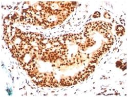
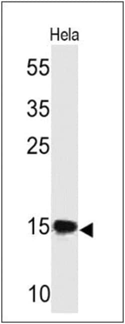
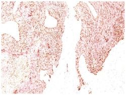
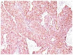
Retinol Binding Protein RBP Mouse, Clone: G4E4, Novus Biologicals™
Mouse Monoclonal Antibody
Manufacturer: Fischer Scientific
The price for this product is unavailable. Please request a quote
Antigen
Retinol Binding Protein RBP
Dilution
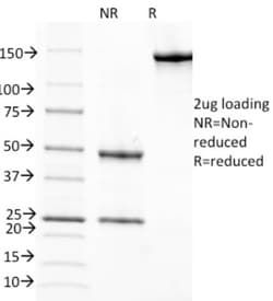
Western Blot 0.5-1ug/ml, Immunocytochemistry/Immunofluorescence 1-2ug/ml, Immunoprecipitation 1-2ug/500ug protein, Immunohistochemistry-Frozen 1-2ug/ml, SDS-Page
Classification
Monoclonal
Form
Purified
Regulatory Status
RUO
Formulation
PBS with 0.05% BSA. with 0.05% Sodium Azide
Gene Alias
C, Cellular retinol-binding protein, CRABP-I, CRBP1CRBP-I, CRBPCellular retinol-binding protein I, CRBPI, RBPC, retinol binding protein 1, cellular, retinol-binding protein 1, retinol-binding protein 1, cellular
Gene Symbols
RBP1
Isotype
IgG1 κ
Purification Method
Protein A purified
Test Specificity
This MAb recognizes an epitope within the 74-182 C-terminal sequence (11kD peptide fragment) of human serum Cellular Retinol Binding Protein 1 (CRBP 1), a single-chain glycoprotein belonging to the superfamily of hydrophobic molecule transporter proteins, which is responsible for transport of retinol (vitamin A1) from the liver to peripheral target tissues, like the eye, where it mediates the cellular uptake. CRBP 1 is synthesized by hepatic parenchymal cells where it becomes bound to its ligand retinol and is then released into the circulation, where it binds further to the protein transthyretin, to form a transporting complex, which is big enough not to be lost by filtration through the kidney glomeruli. It is detected in nearly all tissues with higher expression in adult ovary, pancreas, pituitary gland, adrenal gland, and fetal liver.
Clone
G4E4
Applications
Western Blot, Immunocytochemistry, Immunofluorescence, Immunoprecipitation, Immunohistochemistry (Frozen)
Conjugate
Unconjugated
Host Species
Mouse
Target Species
Human, Mouse, Rat, Goat, Primate, Rabbit
Gene Accession No.
P02753
Gene ID (Entrez)
5947
Immunogen
Retinol binding protein-1 purified from human plasma
Primary or Secondary
Primary
Content And Storage
Store at 4C.
Molecular Weight of Antigen
23 kDa
Related Products
Description
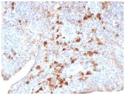
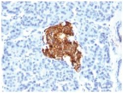
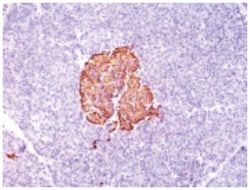
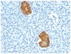
- Retinol Binding Protein RBP Monoclonal specifically detects Retinol Binding Protein RBP in Human, Mouse, Rat, Goat, Chimpanzee, Monkey, Rabbit samples
- It is validated for Immunohistochemistry, Immunohistochemistry-Paraffin.
Compare Similar Items
Show Difference
Antigen: Retinol Binding Protein RBP
Dilution: Western Blot 0.5-1ug/ml, Immunocytochemistry/Immunofluorescence 1-2ug/ml, Immunoprecipitation 1-2ug/500ug protein, Immunohistochemistry-Frozen 1-2ug/ml, SDS-Page
Classification: Monoclonal
Form: Purified
Regulatory Status: RUO
Formulation: PBS with 0.05% BSA. with 0.05% Sodium Azide
Gene Alias: C, Cellular retinol-binding protein, CRABP-I, CRBP1CRBP-I, CRBPCellular retinol-binding protein I, CRBPI, RBPC, retinol binding protein 1, cellular, retinol-binding protein 1, retinol-binding protein 1, cellular
Gene Symbols: RBP1
Isotype: IgG1 κ
Purification Method: Protein A purified
Test Specificity: This MAb recognizes an epitope within the 74-182 C-terminal sequence (11kD peptide fragment) of human serum Cellular Retinol Binding Protein 1 (CRBP 1), a single-chain glycoprotein belonging to the superfamily of hydrophobic molecule transporter proteins, which is responsible for transport of retinol (vitamin A1) from the liver to peripheral target tissues, like the eye, where it mediates the cellular uptake. CRBP 1 is synthesized by hepatic parenchymal cells where it becomes bound to its ligand retinol and is then released into the circulation, where it binds further to the protein transthyretin, to form a transporting complex, which is big enough not to be lost by filtration through the kidney glomeruli. It is detected in nearly all tissues with higher expression in adult ovary, pancreas, pituitary gland, adrenal gland, and fetal liver.
Clone: G4E4
Applications: Western Blot, Immunocytochemistry, Immunofluorescence, Immunoprecipitation, Immunohistochemistry (Frozen)
Conjugate: Unconjugated
Host Species: Mouse
Target Species: Human, Mouse, Rat, Goat, Primate, Rabbit
Gene Accession No.: P02753
Gene ID (Entrez): 5947
Immunogen: Retinol binding protein-1 purified from human plasma
Primary or Secondary: Primary
Content And Storage: Store at 4C.
Molecular Weight of Antigen: 23 kDa

Antigen: Cytokeratin 6
Dilution: Western Blot 0.5-1.0ug/ml, Flow Cytometry 0.5-1ug/million cells, Immunocytochemistry/Immunofluorescence 0.5-1ug/ml, Immunoprecipitation 0.5-1ug/500ug protein lysate, Immunohistochemistry-Paraffin 0.5-1ug/ml, Immunohistochemistry-Frozen 0.5-1ug/ml
Classification: Monoclonal
Form: Purified
Regulatory Status: RUO
Formulation: PBS with 0.05% BSA. with 0.05% Sodium Azide
Gene Alias: Allergen Hom s 5, CK6A, CK-6A, CK6C, CK6D, CK-6D, cytokeratin 6A, cytokeratin 6C, cytokeratin 6D, Cytokeratin-6A, Cytokeratin-6D, K6A, K6C, K6D, keratin 6A, keratin 6C, keratin 6D, keratin, epidermal type II, K6A, keratin, type II cytoskeletal 6A, Keratin-6A, KRT6C, KRT6D, Type-II keratin Kb6
Gene Symbols: KRT6A
Isotype: IgG2a κ
Purification Method: Protein A purified
Test Specificity: This MAb recognizes a protein of 56kDa, identified as cytokeratin 6 (CK6). In humans, multiple isoforms of Cytokeratin 6 (6A-6F), encoded by several highly homologous genes, have distinct tissue expression patterns, and Cytokeratin 6A is the dominant form in epithelial tissue. The gene encoding human Cytokeratin 6A maps to chromosome 12q13, and mutations in this gene are linked to several inheritable hair and skin pathologies. Keratins 6 and 16 are expressed in keratinocytes, which are undergoing rapid turnover in the suprabasal region (also known as hyper-proliferation-related keratins). Keratin 6 is found in hair follicles, suprabasal cells of a variety of internal stratified epithelia, in epidermis, in both normal and hyper-proliferative situations. Epidermal injury results in activation of keratinocytes, which express CK6 and CK16. CK6 is strongly expressed in about 75% of head and neck squamous cell carcinomas. Expression of CK6 is particularly associated with differentiation.
Clone: LHK6 (same as LHK6B)
Applications: Western Blot, Flow Cytometry, Immunocytochemistry, Immunofluorescence, Immunoprecipitation, Immunohistochemistry (Paraffin)
Conjugate: Unconjugated
Host Species: Mouse
Target Species: Human, Mouse
Gene Accession No.: P02538
Gene ID (Entrez): 3853
Immunogen: A synthetic peptide of 11 amino acids (GSSTIKYTTTS) from C-terminus of human keratin 6
Primary or Secondary: Primary
Content And Storage: Store at 4C.
Molecular Weight of Antigen: 56 kDa