Chromogranin A Mouse, Clone: SPM585, Novus Biologicals™
Mouse Monoclonal Antibody
Manufacturer: Fischer Scientific
The price for this product is unavailable. Please request a quote
Antigen
Chromogranin A
Dilution
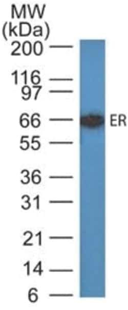
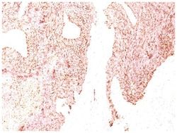
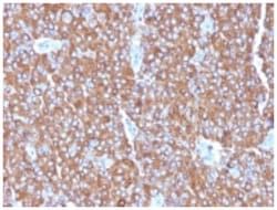
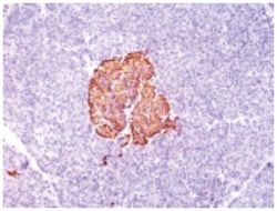
Western Blot 0.5-1ug/ml, Flow Cytometry 0.5-1ug/million cells, Immunocytochemistry/Immunofluorescence 1-2ug/ml, Immunoprecipitation 0.5-1ug/500ug protein lysate, Immunohistochemistry-Paraffin 0.5-1.0ug/ml, Immunohistochemistry-Frozen 0.5-1.0ug/ml
Classification
Monoclonal
Form
Purified
Regulatory Status
RUO
Target Species
Human, Primate, Mouse (Negative), Rat (Negative)
Gene Accession No.
P10645
Gene ID (Entrez)
1113
Immunogen
Human pheochromocytoma
Primary or Secondary
Primary
Content And Storage
Store at 4C.
Clone
SPM585
Applications
Western Blot, Flow Cytometry, Immunocytochemistry, Immunofluorescence, Immunoprecipitation, Immunohistochemistry (Paraffin)
Conjugate
Unconjugated
Host Species
Mouse
Research Discipline
Apoptosis, Cancer, Neuronal Cell Markers
Formulation
PBS with 0.05% BSA. with 0.05% Sodium Azide
Gene Alias
betagranin (N-terminal fragment of chromogranin A), CGA, chromogranin A (parathyroid secretory protein 1), chromogranin-A, parathyroid secretory protein 1, Pituitary secretory protein I, SP-I
Gene Symbols
CHGA
Isotype
IgG1 κ
Purification Method
Protein A purified
Test Specificity
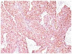
Chromogranin A is present in neuroendocrine cells throughout the body, including the neuroendocrine cells of the large and small intestine, adrenal medulla and pancreatic islets. It is an excellent marker for carcinoid tumors, pheochromocytomas, paragangliomas, and other neuroendocrine tumors. Co-expression of chromogranin A and neuron specific enolase (NSE) is common in neuroendocrine neoplasms. Reportedly, co-expression of certain keratins and chromogranin indicates neuroendocrine lineage. The presence of strong anti-chromogranin staining and absence of anti-keratin staining should raise the possibility of paraganglioma. The co-expression of chromogranin and NSE is typical of neuroendocrine neoplasms. Most pituitary adenomas and prolactinomas readily express chromogranin.
Related Products


Description
- Chromogranin A Monoclonal specifically detects Chromogranin A in Human, Primate, Monkey, Mouse (Negative), Rat (Negative) samples
- It is validated for Flow Cytometry, Immunohistochemistry, Immunocytochemistry/Immunofluorescence, Immunohistochemistry-Paraffin.
Compare Similar Items
Show Difference
Antigen: Chromogranin A
Dilution: Western Blot 0.5-1ug/ml, Flow Cytometry 0.5-1ug/million cells, Immunocytochemistry/Immunofluorescence 1-2ug/ml, Immunoprecipitation 0.5-1ug/500ug protein lysate, Immunohistochemistry-Paraffin 0.5-1.0ug/ml, Immunohistochemistry-Frozen 0.5-1.0ug/ml
Classification: Monoclonal
Form: Purified
Regulatory Status: RUO
Target Species: Human, Primate, Mouse (Negative), Rat (Negative)
Gene Accession No.: P10645
Gene ID (Entrez): 1113
Immunogen: Human pheochromocytoma
Primary or Secondary: Primary
Content And Storage: Store at 4C.
Clone: SPM585
Applications: Western Blot, Flow Cytometry, Immunocytochemistry, Immunofluorescence, Immunoprecipitation, Immunohistochemistry (Paraffin)
Conjugate: Unconjugated
Host Species: Mouse
Research Discipline: Apoptosis, Cancer, Neuronal Cell Markers
Formulation: PBS with 0.05% BSA. with 0.05% Sodium Azide
Gene Alias: betagranin (N-terminal fragment of chromogranin A), CGA, chromogranin A (parathyroid secretory protein 1), chromogranin-A, parathyroid secretory protein 1, Pituitary secretory protein I, SP-I
Gene Symbols: CHGA
Isotype: IgG1 κ
Purification Method: Protein A purified
Test Specificity: Chromogranin A is present in neuroendocrine cells throughout the body, including the neuroendocrine cells of the large and small intestine, adrenal medulla and pancreatic islets. It is an excellent marker for carcinoid tumors, pheochromocytomas, paragangliomas, and other neuroendocrine tumors. Co-expression of chromogranin A and neuron specific enolase (NSE) is common in neuroendocrine neoplasms. Reportedly, co-expression of certain keratins and chromogranin indicates neuroendocrine lineage. The presence of strong anti-chromogranin staining and absence of anti-keratin staining should raise the possibility of paraganglioma. The co-expression of chromogranin and NSE is typical of neuroendocrine neoplasms. Most pituitary adenomas and prolactinomas readily express chromogranin.
Antigen: Cytokeratin 10
Dilution: Western Blot 0.25-0.5ug/ml, Flow Cytometry 0.5-1ug/million cells, Immunocytochemistry/Immunofluorescence 0.5-1ug/ml, Immunoprecipitation 0.5-1ug/500ug protein lysate, Immunohistochemistry-Paraffin 0.5-1.0ug/ml, Immunohistochemistry-Frozen 0.5-1.0ug/ml
Classification: Monoclonal
Form: Purified
Regulatory Status: RUO
Target Species: Human, Mouse
Gene Accession No.: P13645
Gene ID (Entrez): 3858
Immunogen: Skin extract of a human Psoriasis patient
Primary or Secondary: Primary
Content And Storage: Store at 4C.
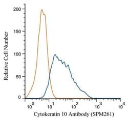
Clone: SPM261
Applications: Western Blot, Flow Cytometry, Immunocytochemistry, Immunofluorescence, Immunoprecipitation, Immunohistochemistry (Paraffin)
Conjugate: Unconjugated
Host Species: Mouse
Research Discipline: Cytoskeleton Markers
Formulation: PBS with 0.05% BSA. with 0.05% Sodium Azide
Gene Alias: BCIE, BIE, CK10, CK-10, cytokeratin 10, Cytokeratin-10, EHK, K10keratosis palmaris et plantaris, keratin 10, Keratin 10 (epidermolytic hyperkeratosis; keratosis palmaris et plantaris), keratin, type I cytoskeletal 10, keratin-10, KPP
Gene Symbols: KRT10
Isotype: IgG1 κ
Purification Method: Protein G purified
Test Specificity: This MAb recognizes a protein of 56.5kDa, identified as cytokeratin 10 (CK10). CK10 is expressed in all suprabasal layers of the epidermis. In the epidermis, expression of CK10 strictly parallels the extent of differentiation; it is absent in the basal layer, appears in the first suprabasal layers and increases in concentration towards the granular layer. However, CK10 is rarely detected in early stages of vulvar squamous carcinomas (tumors less than 2 cm, clinical stage I) regardless of the tumor grade. In larger and more advanced tumors (greater than 2 cm, clinical stages II and III), CK10 is detected very frequently. Expression of CK10 is related to maturation of malignant keratinocytes, being preferentially detected in more-differentiated parts.
Antigen: EPX
Dilution: Flow Cytometry 0.5-1ug/million cells, Immunocytochemistry/Immunofluorescence 0.5-1ug/ml, Immunohistochemistry-Frozen 0.5-1.0ug/ml
Classification: Monoclonal
Form: Purified
Regulatory Status: RUO
Target Species: Human
Gene Accession No.: P11678
Gene ID (Entrez): 8288
Immunogen: Human eosinophils from a patient with hypereosinophilic syndrome
Primary or Secondary: Primary
Content And Storage: Store at 4C.
Clone: AHE-1
Applications: Flow Cytometry, Immunocytochemistry, Immunofluorescence, Immunohistochemistry (Frozen)
Conjugate: Unconjugated
Host Species: Mouse
Research Discipline: __
Formulation: PBS with 0.05% BSA. with 0.05% Sodium Azide
Gene Alias: EC 1.11.1.7, eosinophil peroxidase, EPOEPER, EPPEC 1.11.1, EPX-PEN
Gene Symbols: EPX
Isotype: IgG1 κ
Purification Method: Protein A purified
Test Specificity: Peripheral blood granulocytes are classified into neutrophils, basophils and eosinophils according to the staining characteristics of their cytoplasmic granules. Granule proteins are released by physiologic and pharmacologic stimuli and play important roles in both normal and pathological host immune responses. Eosinophil major basic protein and eosinophil peroxidase (EPX) are granule proteins specific to the eosinophil. AHE-1 recognizes human EPX, a granule protein specific to eosinophils. It does not cross-react with eosinophil major basic protein, elastase, cathepsin G, esterase N, thrombin, plasmin, kallikrein, lactoferrin, or transferrin. This MAb stains eosinophils only and does not stain other peripheral blood cells, including platelets, neutrophils, monocytes, lymphocytes or red blood cells. Human EPX gene product can form a tetramer of two light chains and two heavy chains. Other peroxidase family members include myeloperoxidase (MPO), lactoperoxidase (LPO), and thyroid peroxidase (TPO).