Chromogranin A Mouse, Clone: LK2H10 + PHE5 + CGA/414, Novus Biologicals™
Mouse Monoclonal Antibody
Manufacturer: Fischer Scientific
The price for this product is unavailable. Please request a quote
Antigen
Chromogranin A
Dilution
Western Blot 0.5-1ug/ml, Flow Cytometry 0.5-1ug/million cells, Immunocytochemistry/Immunofluorescence 1-2ug/ml, Immunoprecipitation 1-2ug/500ug protein lysate, Immunohistochemistry-Paraffin 0.5-1ug/ml, Immunohistochemistry-Frozen 0.5-1ug/ml
Classification
Monoclonal
Form
Purified
Regulatory Status
RUO
Target Species
Human, Mouse, Rat, Porcine, Primate
Gene Accession No.
P10645
Gene ID (Entrez)
1113
Immunogen
Human pheochromocytoma (LK2H10 & PHE5) & recombinant human chromogranin A protein (CGA414)
Primary or Secondary
Primary
Content And Storage
Store at 4C.
Molecular Weight of Antigen
72 kDa
Clone
LK2H10 + PHE5 + CGA/414
Applications
Western Blot, Flow Cytometry, Immunocytochemistry, Immunofluorescence, Immunoprecipitation, Immunohistochemistry (Paraffin)
Conjugate
Unconjugated
Host Species
Mouse
Research Discipline
Apoptosis, Cancer, Neuronal Cell Markers
Formulation
PBS with 0.05% BSA. with 0.05% Sodium Azide
Gene Alias
betagranin (N-terminal fragment of chromogranin A), CGA, chromogranin A (parathyroid secretory protein 1), chromogranin-A, parathyroid secretory protein 1, Pituitary secretory protein I, SP-I
Gene Symbols
CHGA
Isotype
IgG
Purification Method
Protein A purified
Test Specificity
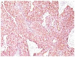
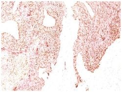
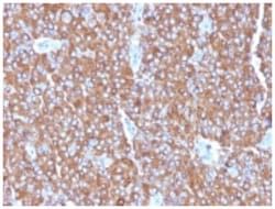
Chromogranin A is present in neuroendocrine cells throughout the body, including the neuroendocrine cells of the large and small intestine, adrenal medulla and pancreatic islets. It is an excellent marker for carcinoid tumors, pheochromocytomas, paragangliomas, and other neuroendocrine tumors. Co-expression of chromogranin A and neuron specific enolase (NSE) is common in neuroendocrine neoplasms. Reportedly, co-expression of certain keratins and chromogranin indicates neuroendocrine lineage. The presence of strong anti-chromogranin staining and absence of anti-keratin staining should raise the possibility of paraganglioma. The co-expression of chromogranin and NSE is typical of neuroendocrine neoplasms. Most pituitary adenomas and prolactinomas readily express chromogranin.
Related Products


Description
- Chromogranin A Monoclonal specifically detects Chromogranin A in Human, Mouse, Rat, Porcine, Monkey samples
- It is validated for Immunohistochemistry, Immunohistochemistry-Paraffin.
Compare Similar Items
Show Difference
Antigen: Chromogranin A
Dilution: Western Blot 0.5-1ug/ml, Flow Cytometry 0.5-1ug/million cells, Immunocytochemistry/Immunofluorescence 1-2ug/ml, Immunoprecipitation 1-2ug/500ug protein lysate, Immunohistochemistry-Paraffin 0.5-1ug/ml, Immunohistochemistry-Frozen 0.5-1ug/ml
Classification: Monoclonal
Form: Purified
Regulatory Status: RUO
Target Species: Human, Mouse, Rat, Porcine, Primate
Gene Accession No.: P10645
Gene ID (Entrez): 1113
Immunogen: Human pheochromocytoma (LK2H10 & PHE5) & recombinant human chromogranin A protein (CGA414)
Primary or Secondary: Primary
Content And Storage: Store at 4C.
Molecular Weight of Antigen: 72 kDa
Clone: LK2H10 + PHE5 + CGA/414
Applications: Western Blot, Flow Cytometry, Immunocytochemistry, Immunofluorescence, Immunoprecipitation, Immunohistochemistry (Paraffin)
Conjugate: Unconjugated
Host Species: Mouse
Research Discipline: Apoptosis, Cancer, Neuronal Cell Markers
Formulation: PBS with 0.05% BSA. with 0.05% Sodium Azide
Gene Alias: betagranin (N-terminal fragment of chromogranin A), CGA, chromogranin A (parathyroid secretory protein 1), chromogranin-A, parathyroid secretory protein 1, Pituitary secretory protein I, SP-I
Gene Symbols: CHGA
Isotype: IgG
Purification Method: Protein A purified
Test Specificity: Chromogranin A is present in neuroendocrine cells throughout the body, including the neuroendocrine cells of the large and small intestine, adrenal medulla and pancreatic islets. It is an excellent marker for carcinoid tumors, pheochromocytomas, paragangliomas, and other neuroendocrine tumors. Co-expression of chromogranin A and neuron specific enolase (NSE) is common in neuroendocrine neoplasms. Reportedly, co-expression of certain keratins and chromogranin indicates neuroendocrine lineage. The presence of strong anti-chromogranin staining and absence of anti-keratin staining should raise the possibility of paraganglioma. The co-expression of chromogranin and NSE is typical of neuroendocrine neoplasms. Most pituitary adenomas and prolactinomas readily express chromogranin.

Antigen: EGFR
Dilution: Western Blot 0.5-1.0ug/ml, Simple Western 10 ug/ml, Immunoprecipitation 1-2ug/500ug protein lysate
Classification: Monoclonal
Form: Purified
Regulatory Status: RUO
Target Species: Human, Mouse (Negative), Rat (Negative)
Gene Accession No.: P00533
Gene ID (Entrez): 1956
Immunogen: Purified EGFR from A431 cells.
Primary or Secondary: Primary
Content And Storage: Store at 4C.
Molecular Weight of Antigen: 170 kDa
Clone: H9B4
Applications: Western Blot, Immunoprecipitation
Conjugate: Unconjugated
Host Species: Mouse
Research Discipline: Cancer, Cell Biology, Cell Cycle and Replication, Growth and Development, Hypoxia, Signal Transduction, Tumor Suppressors, Tyrosine Kinases
Formulation: PBS with 0.05% BSA. with 0.05% Sodium Azide
Gene Alias: avian erythroblastic leukemia viral (v-erb-b) oncogene homolog, cell growth inhibiting protein 40, cell proliferation-inducing protein 61, EC 2.7.10, EC 2.7.10.1, epidermal growth factor receptor, epidermal growth factor receptor (avian erythroblastic leukemia viral (v-erb-b)oncogene homolog), ERBB, ErbB1, ERBB1PIG61, HER1, mENA, Proto-oncogene c-ErbB-1, Receptor tyrosine-protein kinase erbB-1
Gene Symbols: EGFR
Isotype: IgG1
Purification Method: Protein G purified
Test Specificity: This MAb reacts with a cytoplasmic domain of EGFR. EGFR is a type I receptor tyrosine kinase with sequence homology to erbB-1, -2, -3 -4 or HER-1, -2, -3 -4. It binds to Epidermal Growth Factor (EGF), Transforming Growth Factor-a (TGF-a), Heparin-binding EGF (HB-EGF), amphiregulin, beta cellulin & epiregulin.