Cytokeratin 10 Mouse, Clone: SPM261, Novus Biologicals™
Mouse Monoclonal Antibody
Manufacturer: Fischer Scientific
The price for this product is unavailable. Please request a quote
Antigen
Cytokeratin 10
Dilution
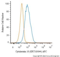
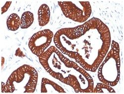
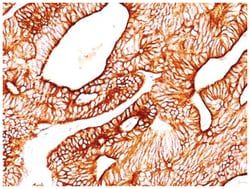
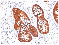
Western Blot 0.25-0.5ug/ml, Flow Cytometry 0.5-1ug/million cells, Immunocytochemistry/Immunofluorescence 0.5-1ug/ml, Immunoprecipitation 0.5-1ug/500ug protein lysate, Immunohistochemistry-Paraffin 0.5-1.0ug/ml, Immunohistochemistry-Frozen 0.5-1.0ug/ml
Classification
Monoclonal
Form
Purified
Regulatory Status
RUO
Target Species
Human, Mouse
Gene Accession No.
P13645
Gene ID (Entrez)
3858
Immunogen
Skin extract of a human Psoriasis patient
Primary or Secondary
Primary
Content And Storage
Store at 4C.
Molecular Weight of Antigen
56.5 kDa
Clone
SPM261
Applications
Western Blot, Flow Cytometry, Immunocytochemistry, Immunofluorescence, Immunoprecipitation, Immunohistochemistry (Paraffin)
Conjugate
Unconjugated
Host Species
Mouse
Research Discipline
Cytoskeleton Markers
Formulation
PBS with 0.05% BSA. with 0.05% Sodium Azide
Gene Alias
BCIE, BIE, CK10, CK-10, cytokeratin 10, Cytokeratin-10, EHK, K10keratosis palmaris et plantaris, keratin 10, Keratin 10 (epidermolytic hyperkeratosis; keratosis palmaris et plantaris), keratin, type I cytoskeletal 10, keratin-10, KPP
Gene Symbols
KRT10
Isotype
IgG1 κ
Purification Method
Protein G purified
Test Specificity
This MAb recognizes a protein of 56.5kDa, identified as cytokeratin 10 (CK10). CK10 is expressed in all suprabasal layers of the epidermis. In the epidermis, expression of CK10 strictly parallels the extent of differentiation; it is absent in the basal layer, appears in the first suprabasal layers and increases in concentration towards the granular layer. However, CK10 is rarely detected in early stages of vulvar squamous carcinomas (tumors less than 2 cm, clinical stage I) regardless of the tumor grade. In larger and more advanced tumors (greater than 2 cm, clinical stages II and III), CK10 is detected very frequently. Expression of CK10 is related to maturation of malignant keratinocytes, being preferentially detected in more-differentiated parts.
Related Products


Description
- Cytokeratin 10 Monoclonal specifically detects Cytokeratin 10 in Human, Mouse samples
- It is validated for Flow Cytometry, Immunohistochemistry, Immunohistochemistry-Paraffin.
Compare Similar Items
Show Difference
Antigen: Cytokeratin 10
Dilution: Western Blot 0.25-0.5ug/ml, Flow Cytometry 0.5-1ug/million cells, Immunocytochemistry/Immunofluorescence 0.5-1ug/ml, Immunoprecipitation 0.5-1ug/500ug protein lysate, Immunohistochemistry-Paraffin 0.5-1.0ug/ml, Immunohistochemistry-Frozen 0.5-1.0ug/ml
Classification: Monoclonal
Form: Purified
Regulatory Status: RUO
Target Species: Human, Mouse
Gene Accession No.: P13645
Gene ID (Entrez): 3858
Immunogen: Skin extract of a human Psoriasis patient
Primary or Secondary: Primary
Content And Storage: Store at 4C.
Molecular Weight of Antigen: 56.5 kDa
Clone: SPM261
Applications: Western Blot, Flow Cytometry, Immunocytochemistry, Immunofluorescence, Immunoprecipitation, Immunohistochemistry (Paraffin)
Conjugate: Unconjugated
Host Species: Mouse
Research Discipline: Cytoskeleton Markers
Formulation: PBS with 0.05% BSA. with 0.05% Sodium Azide
Gene Alias: BCIE, BIE, CK10, CK-10, cytokeratin 10, Cytokeratin-10, EHK, K10keratosis palmaris et plantaris, keratin 10, Keratin 10 (epidermolytic hyperkeratosis; keratosis palmaris et plantaris), keratin, type I cytoskeletal 10, keratin-10, KPP
Gene Symbols: KRT10
Isotype: IgG1 κ
Purification Method: Protein G purified
Test Specificity: This MAb recognizes a protein of 56.5kDa, identified as cytokeratin 10 (CK10). CK10 is expressed in all suprabasal layers of the epidermis. In the epidermis, expression of CK10 strictly parallels the extent of differentiation; it is absent in the basal layer, appears in the first suprabasal layers and increases in concentration towards the granular layer. However, CK10 is rarely detected in early stages of vulvar squamous carcinomas (tumors less than 2 cm, clinical stage I) regardless of the tumor grade. In larger and more advanced tumors (greater than 2 cm, clinical stages II and III), CK10 is detected very frequently. Expression of CK10 is related to maturation of malignant keratinocytes, being preferentially detected in more-differentiated parts.
Antigen: EPX
Dilution: Flow Cytometry 0.5-1ug/million cells, Immunocytochemistry/Immunofluorescence 0.5-1ug/ml, Immunohistochemistry-Frozen 0.5-1.0ug/ml
Classification: Monoclonal
Form: Purified
Regulatory Status: RUO
Target Species: Human
Gene Accession No.: P11678
Gene ID (Entrez): 8288
Immunogen: Human eosinophils from a patient with hypereosinophilic syndrome
Primary or Secondary: Primary
Content And Storage: Store at 4C.
Molecular Weight of Antigen: __
Clone: AHE-1
Applications: Flow Cytometry, Immunocytochemistry, Immunofluorescence, Immunohistochemistry (Frozen)
Conjugate: Unconjugated
Host Species: Mouse
Research Discipline: __
Formulation: PBS with 0.05% BSA. with 0.05% Sodium Azide
Gene Alias: EC 1.11.1.7, eosinophil peroxidase, EPOEPER, EPPEC 1.11.1, EPX-PEN
Gene Symbols: EPX
Isotype: IgG1 κ
Purification Method: Protein A purified
Test Specificity: Peripheral blood granulocytes are classified into neutrophils, basophils and eosinophils according to the staining characteristics of their cytoplasmic granules. Granule proteins are released by physiologic and pharmacologic stimuli and play important roles in both normal and pathological host immune responses. Eosinophil major basic protein and eosinophil peroxidase (EPX) are granule proteins specific to the eosinophil. AHE-1 recognizes human EPX, a granule protein specific to eosinophils. It does not cross-react with eosinophil major basic protein, elastase, cathepsin G, esterase N, thrombin, plasmin, kallikrein, lactoferrin, or transferrin. This MAb stains eosinophils only and does not stain other peripheral blood cells, including platelets, neutrophils, monocytes, lymphocytes or red blood cells. Human EPX gene product can form a tetramer of two light chains and two heavy chains. Other peroxidase family members include myeloperoxidase (MPO), lactoperoxidase (LPO), and thyroid peroxidase (TPO).
Antigen: ER alpha/NR3A1
Dilution: Western Blot 0.5-1ug/ml, Immunohistochemistry-Paraffin 0.5-1.0ug/ml
Classification: Monoclonal
Form: Purified
Regulatory Status: RUO
Target Species: Human
Gene Accession No.: P03372
Gene ID (Entrez): 2099
Immunogen: Recombinant human Estrogen Receptor alpha protein (aa2-185)
Primary or Secondary: Primary
Content And Storage: Store at 4C.
Molecular Weight of Antigen: 67 kDa
Clone: SPM567
Applications: Western Blot, Immunohistochemistry (Paraffin)
Conjugate: Unconjugated
Host Species: Mouse
Research Discipline: Angiogenesis, Breast Cancer, Cancer, Cell Biology, Cell Cycle and Replication, Dendritic Cell Markers, GPCR, Neuroscience, Phospho Specific, Signal Transduction, Transcription Factors and Regulators
Formulation: PBS with 0.05% BSA. with 0.05% Sodium Azide
Gene Alias: ER, ER alpha, Era, ER-alpha, ESRESRA, Estradiol receptor, estrogen receptor, estrogen receptor 1, estrogen receptor alpha, estrogen receptor alpha delta 3*4,56,7*/819-2 isoform, estrogen receptor alpha delta 4 +49 isoform, estrogen receptor alpha delta 4*5,6,7*/654 isoform, NR3A1DKFZp686N23123, Nuclear receptor subfamily 3 group A member 1
Gene Symbols: ESR1
Isotype: IgG1 κ
Purification Method: Protein A purified
Test Specificity: This MAb is specific to ER alpha and shows minimal cross-reaction with other members of the family. ER is an important regulator of growth and differentiation in the mammary gland. Presence of ER in breast tumors indicates an increased likelihood of response to anti-estrogen (e.g. tamoxifen) therapy. This MAb is excellent for staining of formalin-fixed, paraffin-embedded breast carcinomas.