Cytokeratin 6 Mouse, Clone: LHK6 (same as LHK6B), Novus Biologicals™
Mouse Monoclonal Antibody
Manufacturer: Fischer Scientific
The price for this product is unavailable. Please request a quote
Antigen
Cytokeratin 6
Dilution
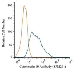
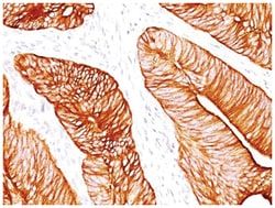
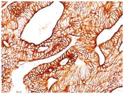
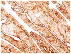
Western Blot 0.5-1.0ug/ml, Flow Cytometry 0.5-1ug/million cells, Immunocytochemistry/Immunofluorescence 0.5-1ug/ml, Immunoprecipitation 0.5-1ug/500ug protein lysate, Immunohistochemistry-Paraffin 0.5-1ug/ml, Immunohistochemistry-Frozen 0.5-1ug/ml
Classification
Monoclonal
Form
Purified
Regulatory Status
RUO
Target Species
Human, Mouse
Gene Accession No.
P02538
Gene ID (Entrez)
3853
Immunogen
A synthetic peptide of 11 amino acids (GSSTIKYTTTS) from C-terminus of human keratin 6
Primary or Secondary
Primary
Content And Storage
Store at 4C.
Molecular Weight of Antigen
56 kDa
Clone
LHK6 (same as LHK6B)
Applications
Western Blot, Flow Cytometry, Immunocytochemistry, Immunofluorescence, Immunoprecipitation, Immunohistochemistry (Paraffin)
Conjugate
Unconjugated
Host Species
Mouse
Research Discipline
Cytoskeleton Markers, Stem Cell Markers
Formulation
PBS with 0.05% BSA. with 0.05% Sodium Azide
Gene Alias
Allergen Hom s 5, CK6A, CK-6A, CK6C, CK6D, CK-6D, cytokeratin 6A, cytokeratin 6C, cytokeratin 6D, Cytokeratin-6A, Cytokeratin-6D, K6A, K6C, K6D, keratin 6A, keratin 6C, keratin 6D, keratin, epidermal type II, K6A, keratin, type II cytoskeletal 6A, Keratin-6A, KRT6C, KRT6D, Type-II keratin Kb6
Gene Symbols
KRT6A
Isotype
IgG2a κ
Purification Method
Protein A purified
Test Specificity
This MAb recognizes a protein of 56kDa, identified as cytokeratin 6 (CK6). In humans, multiple isoforms of Cytokeratin 6 (6A-6F), encoded by several highly homologous genes, have distinct tissue expression patterns, and Cytokeratin 6A is the dominant form in epithelial tissue. The gene encoding human Cytokeratin 6A maps to chromosome 12q13, and mutations in this gene are linked to several inheritable hair and skin pathologies. Keratins 6 and 16 are expressed in keratinocytes, which are undergoing rapid turnover in the suprabasal region (also known as hyper-proliferation-related keratins). Keratin 6 is found in hair follicles, suprabasal cells of a variety of internal stratified epithelia, in epidermis, in both normal and hyper-proliferative situations. Epidermal injury results in activation of keratinocytes, which express CK6 and CK16. CK6 is strongly expressed in about 75% of head and neck squamous cell carcinomas. Expression of CK6 is particularly associated with differentiation.
Related Products


Description
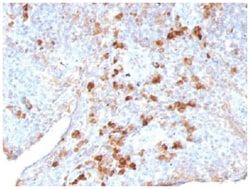
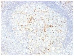
- Cytokeratin 6 Monoclonal specifically detects Cytokeratin 6 in Human, Mouse samples
- It is validated for Flow Cytometry, Immunohistochemistry, Immunocytochemistry/Immunofluorescence, Immunohistochemistry-Paraffin.
Compare Similar Items
Show Difference
Antigen: Cytokeratin 6
Dilution: Western Blot 0.5-1.0ug/ml, Flow Cytometry 0.5-1ug/million cells, Immunocytochemistry/Immunofluorescence 0.5-1ug/ml, Immunoprecipitation 0.5-1ug/500ug protein lysate, Immunohistochemistry-Paraffin 0.5-1ug/ml, Immunohistochemistry-Frozen 0.5-1ug/ml
Classification: Monoclonal
Form: Purified
Regulatory Status: RUO
Target Species: Human, Mouse
Gene Accession No.: P02538
Gene ID (Entrez): 3853
Immunogen: A synthetic peptide of 11 amino acids (GSSTIKYTTTS) from C-terminus of human keratin 6
Primary or Secondary: Primary
Content And Storage: Store at 4C.
Molecular Weight of Antigen: 56 kDa
Clone: LHK6 (same as LHK6B)
Applications: Western Blot, Flow Cytometry, Immunocytochemistry, Immunofluorescence, Immunoprecipitation, Immunohistochemistry (Paraffin)
Conjugate: Unconjugated
Host Species: Mouse
Research Discipline: Cytoskeleton Markers, Stem Cell Markers
Formulation: PBS with 0.05% BSA. with 0.05% Sodium Azide
Gene Alias: Allergen Hom s 5, CK6A, CK-6A, CK6C, CK6D, CK-6D, cytokeratin 6A, cytokeratin 6C, cytokeratin 6D, Cytokeratin-6A, Cytokeratin-6D, K6A, K6C, K6D, keratin 6A, keratin 6C, keratin 6D, keratin, epidermal type II, K6A, keratin, type II cytoskeletal 6A, Keratin-6A, KRT6C, KRT6D, Type-II keratin Kb6
Gene Symbols: KRT6A
Isotype: IgG2a κ
Purification Method: Protein A purified
Test Specificity: This MAb recognizes a protein of 56kDa, identified as cytokeratin 6 (CK6). In humans, multiple isoforms of Cytokeratin 6 (6A-6F), encoded by several highly homologous genes, have distinct tissue expression patterns, and Cytokeratin 6A is the dominant form in epithelial tissue. The gene encoding human Cytokeratin 6A maps to chromosome 12q13, and mutations in this gene are linked to several inheritable hair and skin pathologies. Keratins 6 and 16 are expressed in keratinocytes, which are undergoing rapid turnover in the suprabasal region (also known as hyper-proliferation-related keratins). Keratin 6 is found in hair follicles, suprabasal cells of a variety of internal stratified epithelia, in epidermis, in both normal and hyper-proliferative situations. Epidermal injury results in activation of keratinocytes, which express CK6 and CK16. CK6 is strongly expressed in about 75% of head and neck squamous cell carcinomas. Expression of CK6 is particularly associated with differentiation.
Antigen: Lambda Light Chain
Dilution: Western Blot 0.5-1.0ug/ml, Flow Cytometry 0.5-1ug/million cells, Immunocytochemistry/Immunofluorescence 0.5-1ug/ml, Immunoprecipitation 0.5-1ug/500ug protein lysate, Immunohistochemistry-Paraffin 0.5-1ug/ml, Immunohistochemistry-Frozen 0.5-1ug/ml, SDS-Page
Classification: Monoclonal
Form: Purified
Regulatory Status: RUO
Target Species: Human
Gene Accession No.: P01701
Gene ID (Entrez): 3536
Immunogen: Purified human lambda light chain
Primary or Secondary: Primary
Content And Storage: Store at 4C.
Molecular Weight of Antigen: 22.5 kDa
Clone: LAM03
Applications: Western Blot, Flow Cytometry, Immunocytochemistry, Immunofluorescence, Immunoprecipitation, Immunohistochemistry (Paraffin)
Conjugate: Unconjugated
Host Species: Mouse
Research Discipline: __
Formulation: PBS with 0.05% BSA. with 0.05% Sodium Azide
Gene Alias: IGLC, immunoglobulin lambda constant group
Gene Symbols: IGL
Isotype: IgG1 κ
Purification Method: Protein A purified
Test Specificity: This MAb is specific to lambda light chain of immunoglobulin and shows no cross-reaction with lambda light chain or any of the five heavy chains. In mammals, the two light chains in an antibody are always identical, with only one type of light chain, kappa or lambda. The ratio of Kappa to Lambda is 70:30. However, with the occurrence of multiple myeloma or other B-cell malignancies this ratio is disturbed. Antibody to the lambda light chain is reportedly useful in the identification of leukemias, plasmacytomas, and certain non-Hodgkin's lymphomas. Demonstration of clonality in lymphoid infiltrates indicates that the infiltrate is malignant.
Antigen: Lambda Light Chain
Dilution: Western Blot 0.5-1.0ug/ml, Flow Cytometry 0.5-1ug/million cells, Immunocytochemistry/Immunofluorescence 0.5-1ug/ml, Immunoprecipitation 0.5-1ug/500ug protein lysate, Immunohistochemistry-Paraffin 0.5-1ug/ml, Immunohistochemistry-Frozen 0.5-1ug/ml
Classification: Monoclonal
Form: Purified
Regulatory Status: RUO
Target Species: Human
Gene Accession No.: P01701
Gene ID (Entrez): 3536
Immunogen: Purified human IgG (LcN-2 and ICO-106)
Primary or Secondary: Primary
Content And Storage: Store at 4C.
Molecular Weight of Antigen: 22.5 kDa
Clone: LcN-2 + ICO-106
Applications: Western Blot, Flow Cytometry, Immunocytochemistry, Immunofluorescence, Immunoprecipitation, Immunohistochemistry (Paraffin)
Conjugate: Unconjugated
Host Species: Mouse
Research Discipline: __
Formulation: PBS with 0.05% BSA. with 0.05% Sodium Azide
Gene Alias: IGLC, immunoglobulin lambda constant group
Gene Symbols: IGL
Isotype: IgG1, IgG2a κ
Purification Method: Protein A purified
Test Specificity: This MAb is specific to lambda light chain of immunoglobulin and shows no cross-reaction with lambda light chain or any of the five heavy chains. In mammals, the two light chains in an antibody are always identical, with only one type of light chain, kappa or lambda. The ratio of Kappa to Lambda is 70:30. However, with the occurrence of multiple myeloma or other B-cell malignancies this ratio is disturbed. Antibody to the lambda light chain is reportedly useful in the identification of leukemias, plasmacytomas, and certain non-Hodgkin's lymphomas. Demonstration of clonality in lymphoid infiltrates indicates that the infiltrate is malignant.