Melan-A/MART-1 Mouse, Clone: SPM540, Novus Biologicals™
Manufacturer: Fischer Scientific
The price for this product is unavailable. Please request a quote
Antigen
Melan-A/MART-1
Classification
Monoclonal
Conjugate
Unconjugated
Formulation
PBS with 0.05% BSA. with 0.05% Sodium Azide
Gene Alias
Antigen LB39-AA, Antigen SK29-AA, Mart 1 Melan A, MART1MART-1, melan-A, melanoma antigen recognized by T-cells 1, Protein Melan-A
Host Species
Mouse
Purification Method
Protein A purified
Regulatory Status
RUO
Primary or Secondary
Primary
Test Specificity
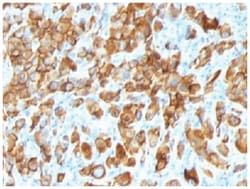
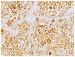
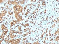
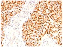
This antibody recognizes a protein doublet of 20-22kDa, identified as MART-1 (Melanoma Antigen Recognized by T cells 1) or Melan-A. MART-1 is a newly identified melanocyte differentiation antigen recognized by autologous cytotoxic T lymphocytes. Seven other melanoma associated antigens recognized by autologous cytotoxic T cells include MAGE-1, MAGE-3, tyrosinase, gp100, gp75, BAGE-1, and GAGE-1. Subcellular fractionation shows that MART-1 is present in melanosomes and endoplasmic reticulum. This MAb labels melanomas and other tumors showing melanocytic differentiation. It is also a useful positive-marker for angiomyolipomas. It does not stain tumor cells of epithelial, lymphoid, glial, or mesenchymal origin.
Content And Storage
Store at 4C.
Isotype
IgG2b κ
Applications
Western Blot, Flow Cytometry, Immunocytochemistry, Immunofluorescence, Immunoprecipitation, Immunohistochemistry (Paraffin)
Clone
SPM540
Dilution
Western Blot 0.5-1ug/ml, Flow Cytometry 0.5-1ug/million cells, Immunocytochemistry/Immunofluorescence 1-2ug/ml, Immunoprecipitation 0.5-1ug/500ug protein lysate, Immunohistochemistry-Paraffin 0.5-1.0ug/ml, Immunohistochemistry-Frozen 0.5-1.0ug/ml
Gene Accession No.
Q16655
Gene Symbols
MLANA
Immunogen
Recombinant hMART-1 protein
Quantity
0.2 mg
Research Discipline
Cytoskeleton Markers, Immunology
Gene ID (Entrez)
2315
Target Species
Human, Mouse, Rat
Form
Purified
Related Products

Description
- Melan-A/MART-1 Monoclonal specifically detects Melan-A/MART-1 in Human, Mouse, Rat samples
- It is validated for Flow Cytometry, Immunohistochemistry, Immunocytochemistry/Immunofluorescence, Immunohistochemistry-Paraffin.
Compare Similar Items
Show Difference
Antigen: Melan-A/MART-1
Classification: Monoclonal
Conjugate: Unconjugated
Formulation: PBS with 0.05% BSA. with 0.05% Sodium Azide
Gene Alias: Antigen LB39-AA, Antigen SK29-AA, Mart 1 Melan A, MART1MART-1, melan-A, melanoma antigen recognized by T-cells 1, Protein Melan-A
Host Species: Mouse
Purification Method: Protein A purified
Regulatory Status: RUO
Primary or Secondary: Primary
Test Specificity: This antibody recognizes a protein doublet of 20-22kDa, identified as MART-1 (Melanoma Antigen Recognized by T cells 1) or Melan-A. MART-1 is a newly identified melanocyte differentiation antigen recognized by autologous cytotoxic T lymphocytes. Seven other melanoma associated antigens recognized by autologous cytotoxic T cells include MAGE-1, MAGE-3, tyrosinase, gp100, gp75, BAGE-1, and GAGE-1. Subcellular fractionation shows that MART-1 is present in melanosomes and endoplasmic reticulum. This MAb labels melanomas and other tumors showing melanocytic differentiation. It is also a useful positive-marker for angiomyolipomas. It does not stain tumor cells of epithelial, lymphoid, glial, or mesenchymal origin.
Content And Storage: Store at 4C.
Isotype: IgG2b κ
Applications: Western Blot, Flow Cytometry, Immunocytochemistry, Immunofluorescence, Immunoprecipitation, Immunohistochemistry (Paraffin)
Clone: SPM540
Dilution: Western Blot 0.5-1ug/ml, Flow Cytometry 0.5-1ug/million cells, Immunocytochemistry/Immunofluorescence 1-2ug/ml, Immunoprecipitation 0.5-1ug/500ug protein lysate, Immunohistochemistry-Paraffin 0.5-1.0ug/ml, Immunohistochemistry-Frozen 0.5-1.0ug/ml
Gene Accession No.: Q16655
Gene Symbols: MLANA
Immunogen: Recombinant hMART-1 protein
Quantity: 0.2 mg
Research Discipline: Cytoskeleton Markers, Immunology
Gene ID (Entrez): 2315
Target Species: Human, Mouse, Rat
Form: Purified
Antigen: Melan-A/MART-1
Classification: Monoclonal
Conjugate: Unconjugated
Formulation: PBS with 0.05% BSA. with 0.05% Sodium Azide
Gene Alias: Antigen LB39-AA, Antigen SK29-AA, Mart 1 Melan A, MART1MART-1, melan-A, melanoma antigen recognized by T-cells 1, Protein Melan-A
Host Species: Mouse
Purification Method: Protein A purified
Regulatory Status: RUO
Primary or Secondary: Primary
Test Specificity: This antibody recognizes a protein doublet of 20-22kDa, identified as MART-1 (Melanoma Antigen Recognized by T cells 1) or Melan-A. MART-1 is a newly identified melanocyte differentiation antigen recognized by autologous cytotoxic T lymphocytes. Seven other melanoma associated antigens recognized by autologous cytotoxic T cells include MAGE-1, MAGE-3, tyrosinase, gp100, gp75, BAGE-1, and GAGE-1. Subcellular fractionation shows that MART-1 is present in melanosomes and endoplasmic reticulum. This MAb labels melanomas and other tumors showing melanocytic differentiation. It is also a useful positive-marker for angiomyolipomas. It does not stain tumor cells of epithelial, lymphoid, glial, or mesenchymal origin.
Content And Storage: Store at 4C.
Isotype: IgG2b κ
Applications: Western Blot, Flow Cytometry, Immunocytochemistry, Immunofluorescence, Immunoprecipitation, Immunohistochemistry (Paraffin)
Clone: SPM540
Dilution: Western Blot 0.5-1ug/ml, Flow Cytometry 0.5-1ug/million cells, Immunocytochemistry/Immunofluorescence 1-2ug/ml, Immunoprecipitation 0.5-1ug/500ug protein lysate, Immunohistochemistry-Paraffin 0.5-1.0ug/ml, Immunohistochemistry-Frozen 0.5-1.0ug/ml
Gene Accession No.: Q16655
Gene Symbols: MLANA
Immunogen: Recombinant hMART-1 protein
Quantity: 0.02 mg
Research Discipline: Cytoskeleton Markers, Immunology
Gene ID (Entrez): 2315
Target Species: Human, Mouse, Rat
Form: Purified
Antigen: MUC2
Classification: Monoclonal
Conjugate: Unconjugated
Formulation: PBS with 0.05% BSA. with 0.05% Sodium Azide
Gene Alias: Intestinal mucin-2, MLP, MUC-2, mucin 2, intestinal/tracheal, mucin 2, oligomeric mucus/gel-forming, mucin-2, mucin-like protein, SMUC
Host Species: Mouse
Purification Method: Protein A purified
Regulatory Status: RUO
Primary or Secondary: Primary
Test Specificity: Recognizes a single glycoprotein of 520kDa, identified as mucin 2 (MUC2). This MAb shows no cross-reaction with human milk fat globule membranes, MUC1, or MUC3. Mucins are high molecular weight glycoproteins, which constitute the major component of the mucus layer that protects the gastric epithelium.MUC2 is specifically expressed in goblet cells of the small intestine & colon; in about 65% of colonic carcinomas and about 40% of gastric carcinomas. MUC2 is rarely expressed outside of the GI tract with the exceptions of mucinous carcinoma of breast and clear cell-type carcinomas of the ovary.
Content And Storage: Store at 4C.
Isotype: IgG1 κ
Applications: Western Blot, Flow Cytometry, Immunocytochemistry, Immunofluorescence, Immunoprecipitation, Immunohistochemistry (Paraffin)
Clone: SPM296
Dilution: Western Blot 0.5-1ug/ml, Flow Cytometry 0.5-1ug/million cells, Immunocytochemistry/Immunofluorescence 1-2ug/ml, Immunoprecipitation 0.5-1ug/500ug protein lysate, Immunohistochemistry-Paraffin 0.5-1.0ug/ml, Immunohistochemistry-Frozen 0.5-1.0ug/ml
Gene Accession No.: Q02817
Gene Symbols: MUC2
Immunogen: A synthetic peptide of 29 amino acids KYPTTTPISTTTMVTPTPTPTGTQTPTTT from MUC2 protein, coupled to KLH.
Quantity: 0.1 mg
Research Discipline: __
Gene ID (Entrez): 4583
Target Species: Human
Form: Purified