Melan-A/MART-1 Antibody (MLANA/788), Novus Biologicals™
Manufacturer: Fischer Scientific
Select a Size
| Pack Size | SKU | Availability | Price |
|---|---|---|---|
| Each of 1 | NBP24439401-Each-of-1 | In Stock | ₹ 46,636.00 |
NBP24439401 - Each of 1
In Stock
Quantity
1
Base Price: ₹ 46,636.00
GST (18%): ₹ 8,394.48
Total Price: ₹ 55,030.48
Antigen
Melan-A/MART-1
Classification
Monoclonal
Concentration
0.2mg/mL
Dilution
Western Blot 0.5 - 1.0 ug/ml, Flow Cytometry 0.5 - 1 ug/million cells in 0.1 ml, Immunohistochemistry-Paraffin 0.5 - 1.0 ug/ml, Immunofluorescence 0.5 - 1.0 ug/ml
Gene Accession No.
Q16655
Gene Symbols
MLANA
Immunogen
Recombinant human MLANA protein
Quantity
0.1 mg
Research Discipline
Cytoskeleton Markers, Immunology
Gene ID (Entrez)
2315
Target Species
Human, Mouse, Rat, Canine
Form
Purified
Applications
Western Blot, Flow Cytometry, Immunohistochemistry (Paraffin), Immunofluorescence
Clone
MLANA/788
Conjugate
Unconjugated
Formulation
10mM PBS and 0.05% BSA with 0.05% Sodium Azide
Gene Alias
Antigen LB39-AA, Antigen SK29-AA, Mart 1 Melan A, MART1MART-1, melan-A, melanoma antigen recognized by T-cells 1, Protein Melan-A
Host Species
Mouse
Purification Method
Protein A or G purified
Regulatory Status
RUO
Primary or Secondary
Primary
Test Specificity
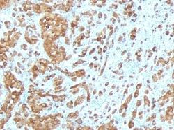
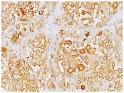
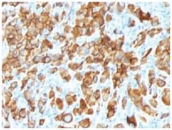
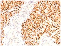
This antibody recognizes a protein doublet of 20-22kDa, identified as MART-1 (Melanoma Antigen Recognized by T cells 1) or Melan-A. MART-1 is a newly identified melanocyte differentiation antigen recognized by autologous cytotoxic T lymphocytes. Seven other melanoma associated antigens recognized by autologous cytotoxic T cells include MAGE-1, MAGE-3, tyrosinase, gp100, gp75, BAGE-1, and GAGE-1. Subcellular fractionation shows that MART-1 is present in melanosomes and endoplasmic reticulum. This MAb labels melanomas and other tumors showing melanocytic differentiation. It is also a useful positive-marker for angiomyolipomas. It does not stain tumor cells of epithelial, lymphoid, glial, or mesenchymal origin.
Content And Storage
Store at 4C.
Isotype
IgG1 κ
Related Products

Description
- Ensure accurate, reproducible results in Western Blot, Flow Cytometry, Immunohistochemistry (Paraffin), Immunofluorescence Melan-A/MART-1 Monoclonal specifically detects Melan-A/MART-1 in Human, Mouse, Rat, Canine samples
- It is validated for Western Blot, Flow Cytometry, Immunohistochemistry, Immunohistochemistry-Paraffin.
Compare Similar Items
Show Difference
Antigen: Melan-A/MART-1
Classification: Monoclonal
Concentration: 0.2mg/mL
Dilution: Western Blot 0.5 - 1.0 ug/ml, Flow Cytometry 0.5 - 1 ug/million cells in 0.1 ml, Immunohistochemistry-Paraffin 0.5 - 1.0 ug/ml, Immunofluorescence 0.5 - 1.0 ug/ml
Gene Accession No.: Q16655
Gene Symbols: MLANA
Immunogen: Recombinant human MLANA protein
Quantity: 0.1 mg
Research Discipline: Cytoskeleton Markers, Immunology
Gene ID (Entrez): 2315
Target Species: Human, Mouse, Rat, Canine
Form: Purified
Applications: Western Blot, Flow Cytometry, Immunohistochemistry (Paraffin), Immunofluorescence
Clone: MLANA/788
Conjugate: Unconjugated
Formulation: 10mM PBS and 0.05% BSA with 0.05% Sodium Azide
Gene Alias: Antigen LB39-AA, Antigen SK29-AA, Mart 1 Melan A, MART1MART-1, melan-A, melanoma antigen recognized by T-cells 1, Protein Melan-A
Host Species: Mouse
Purification Method: Protein A or G purified
Regulatory Status: RUO
Primary or Secondary: Primary
Test Specificity: This antibody recognizes a protein doublet of 20-22kDa, identified as MART-1 (Melanoma Antigen Recognized by T cells 1) or Melan-A. MART-1 is a newly identified melanocyte differentiation antigen recognized by autologous cytotoxic T lymphocytes. Seven other melanoma associated antigens recognized by autologous cytotoxic T cells include MAGE-1, MAGE-3, tyrosinase, gp100, gp75, BAGE-1, and GAGE-1. Subcellular fractionation shows that MART-1 is present in melanosomes and endoplasmic reticulum. This MAb labels melanomas and other tumors showing melanocytic differentiation. It is also a useful positive-marker for angiomyolipomas. It does not stain tumor cells of epithelial, lymphoid, glial, or mesenchymal origin.
Content And Storage: Store at 4C.
Isotype: IgG1 κ
Antigen: Melan-A/MART-1
Classification: Monoclonal
Concentration: 0.2mg/mL
Dilution: Western Blot 0.5 - 1.0 ug/ml, Flow Cytometry 0.5 - 1 ug/million cells in 0.1 ml, Immunohistochemistry-Paraffin 0.5 - 1.0 ug/ml, Immunofluorescence 0.5 - 1.0 ug/ml
Gene Accession No.: Q16655
Gene Symbols: MLANA
Immunogen: Recombinant human MLANA protein
Quantity: 0.2 mg
Research Discipline: Cytoskeleton Markers, Immunology
Gene ID (Entrez): 2315
Target Species: Human, Mouse, Rat, Canine
Form: Purified
Applications: Western Blot, Flow Cytometry, Immunohistochemistry (Paraffin), Immunofluorescence
Clone: MLANA/788
Conjugate: Unconjugated
Formulation: 10mM PBS and 0.05% BSA with 0.05% Sodium Azide
Gene Alias: Antigen LB39-AA, Antigen SK29-AA, Mart 1 Melan A, MART1MART-1, melan-A, melanoma antigen recognized by T-cells 1, Protein Melan-A
Host Species: Mouse
Purification Method: Protein A or G purified
Regulatory Status: RUO
Primary or Secondary: Primary
Test Specificity: This antibody recognizes a protein doublet of 20-22kDa, identified as MART-1 (Melanoma Antigen Recognized by T cells 1) or Melan-A. MART-1 is a newly identified melanocyte differentiation antigen recognized by autologous cytotoxic T lymphocytes. Seven other melanoma associated antigens recognized by autologous cytotoxic T cells include MAGE-1, MAGE-3, tyrosinase, gp100, gp75, BAGE-1, and GAGE-1. Subcellular fractionation shows that MART-1 is present in melanosomes and endoplasmic reticulum. This MAb labels melanomas and other tumors showing melanocytic differentiation. It is also a useful positive-marker for angiomyolipomas. It does not stain tumor cells of epithelial, lymphoid, glial, or mesenchymal origin.
Content And Storage: Store at 4C.
Isotype: IgG1 κ
Antigen: ECM-1/Secretory Component P85
Classification: Monoclonal
Concentration: 0.2mg/mL
Dilution: Flow Cytometry 0.5 - 1 ug/million cells in 0.1 ml, Immunohistochemistry-Paraffin 0.5 - 1.0 ug/ml, SDS-Page, Immunofluorescence 1 - 2 ug/ml
Gene Accession No.: Q16610
Gene Symbols: ECM1
Immunogen: Recombinant human ECM1 protein
Quantity: 0.02 mg
Research Discipline: Extracellular Matrix, Immunology
Gene ID (Entrez): 1893
Target Species: Human, Rat
Form: Purified
Applications: Flow Cytometry, Immunohistochemistry (Paraffin), SDS-Page, Immunofluorescence
Clone: ECM1/792
Conjugate: Unconjugated
Formulation: 10mM PBS and 0.05% BSA with 0.05% Sodium Azide
Gene Alias: ECM1, extracellular matrix protein 1, Secretory Component Glycoprotein, Secretory component p85
Host Species: Mouse
Purification Method: Protein A or G purified
Regulatory Status: RUO
Primary or Secondary: Primary
Test Specificity: This MAb reacts with a reduction-resistant epitope present in both free and SIgA bound Secretory Component. It does not react with the cell lines lacking secretory component. The antibody is useful for studying the distribution and level of both free and bound secretory component. Secretory component is differentially expressed in epithelium, and the antibody is a popular marker for identifying subpopulations of epithelial cells and epithelial differentiation. The Secretory component antibody is a useful research tool for studying mucosal immunity, inflammation, remodeling, differentiation and tumorigenesis, all processes associated with differential secretory component expression.
Content And Storage: Store at 4C.
Isotype: IgG1 κ