NF-H Antibody (RT97), Alexa Fluor™ 647, Novus Biologicals™
Manufacturer: Novus Biologicals
Select a Size
| Pack Size | SKU | Availability | Price |
|---|---|---|---|
| Each of 1 | NBP233062Y-Each-of-1 | In Stock | ₹ 57,494.00 |
NBP233062Y - Each of 1
In Stock
Quantity
1
Base Price: ₹ 57,494.00
GST (18%): ₹ 10,348.92
Total Price: ₹ 67,842.92
Antigen
NF-H
Classification
Monoclonal
Conjugate
Alexa Fluor 647
Gene Alias
200 kDa neurofilament protein, KIAA0845, Neurofilament Heavy (200kDa), neurofilament heavy polypeptide, Neurofilament triplet H protein, neurofilament, heavy polypeptide, neurofilament, heavy polypeptide 200kDa, NF200, NFH, NF-H
Host Species
Mouse
Purification Method
Protein A or G purified
Regulatory Status
RUO
Primary or Secondary
Primary
Test Specificity
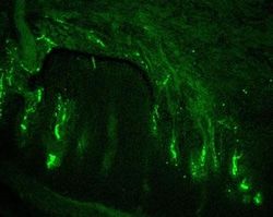
This monoclonal antibody reacts with a 200kDa protein, identified as heavy sub-unit of neurofilaments (NF-H). Neurofilaments make up the main structural elements of axons and dendrites and are found in neurons, peripheral nerves, and sympathetic ganglion cells. Neurofilaments consist of three major subunits with molecular weights of 68kDa (NF-L), 160kDa (NF-M) and 200kDa (NF-H). Anti-neurofilament stains a number of neural, neuroendocrine, and endocrine tumors. Neuromas, ganglioneuromas, gangliogliomas, ganglioneuroblastomas, and neuroblastomas stain positively for anti-neurofilament. Neurofilaments are also present in paragangliomas as well as adrenal and extra-adrenal pheochromocytomas. Carcinoids, neuroendocrine carcinomas of the skin, and oat cell carcinomas of the lung also express neurofilament.
Content And Storage
Store at 4C in the dark.
Isotype
IgG1 κ
Applications
ELISA
Clone
RT97
Dilution
ELISA
Gene Symbols
NEFH
Immunogen
Triton-X 100 insoluble protein fraction of rat brain (Uniprot: P12036)
Quantity
0.1 mL
Research Discipline
Autophagy, Cellular Markers, Cytoskeleton Markers, Neurodegeneration, Neurofilaments, Neuronal Cell Markers, Neuroscience
Gene ID (Entrez)
4744
Target Species
Human, Mouse, Rat, Porcine, Chicken
Form
Purified
Related Products


Description
- NF-H Monoclonal specifically detects NF-H in Human, Mouse, Rat, Porcine, Chicken samples
- It is validated for Western Blot, Flow Cytometry, Immunohistochemistry, Immunohistochemistry-Paraffin.
Compare Similar Items
Show Difference
Antigen: NF-H
Classification: Monoclonal
Conjugate: Alexa Fluor 647
Gene Alias: 200 kDa neurofilament protein, KIAA0845, Neurofilament Heavy (200kDa), neurofilament heavy polypeptide, Neurofilament triplet H protein, neurofilament, heavy polypeptide, neurofilament, heavy polypeptide 200kDa, NF200, NFH, NF-H
Host Species: Mouse
Purification Method: Protein A or G purified
Regulatory Status: RUO
Primary or Secondary: Primary
Test Specificity: This monoclonal antibody reacts with a 200kDa protein, identified as heavy sub-unit of neurofilaments (NF-H). Neurofilaments make up the main structural elements of axons and dendrites and are found in neurons, peripheral nerves, and sympathetic ganglion cells. Neurofilaments consist of three major subunits with molecular weights of 68kDa (NF-L), 160kDa (NF-M) and 200kDa (NF-H). Anti-neurofilament stains a number of neural, neuroendocrine, and endocrine tumors. Neuromas, ganglioneuromas, gangliogliomas, ganglioneuroblastomas, and neuroblastomas stain positively for anti-neurofilament. Neurofilaments are also present in paragangliomas as well as adrenal and extra-adrenal pheochromocytomas. Carcinoids, neuroendocrine carcinomas of the skin, and oat cell carcinomas of the lung also express neurofilament.
Content And Storage: Store at 4C in the dark.
Isotype: IgG1 κ
Applications: ELISA
Clone: RT97
Dilution: ELISA
Gene Symbols: NEFH
Immunogen: Triton-X 100 insoluble protein fraction of rat brain (Uniprot: P12036)
Quantity: 0.1 mL
Research Discipline: Autophagy, Cellular Markers, Cytoskeleton Markers, Neurodegeneration, Neurofilaments, Neuronal Cell Markers, Neuroscience
Gene ID (Entrez): 4744
Target Species: Human, Mouse, Rat, Porcine, Chicken
Form: Purified
Antigen: NF-H
Classification: Monoclonal
Conjugate: Alexa Fluor 700
Gene Alias: 200 kDa neurofilament protein, KIAA0845, Neurofilament Heavy (200kDa), neurofilament heavy polypeptide, Neurofilament triplet H protein, neurofilament, heavy polypeptide, neurofilament, heavy polypeptide 200kDa, NF200, NFH, NF-H
Host Species: Mouse
Purification Method: Protein A or G purified
Regulatory Status: RUO
Primary or Secondary: Primary
Test Specificity: This monoclonal antibody reacts with a 200kDa protein, identified as heavy sub-unit of neurofilaments (NF-H). Neurofilaments make up the main structural elements of axons and dendrites and are found in neurons, peripheral nerves, and sympathetic ganglion cells. Neurofilaments consist of three major subunits with molecular weights of 68kDa (NF-L), 160kDa (NF-M) and 200kDa (NF-H). Anti-neurofilament stains a number of neural, neuroendocrine, and endocrine tumors. Neuromas, ganglioneuromas, gangliogliomas, ganglioneuroblastomas, and neuroblastomas stain positively for anti-neurofilament. Neurofilaments are also present in paragangliomas as well as adrenal and extra-adrenal pheochromocytomas. Carcinoids, neuroendocrine carcinomas of the skin, and oat cell carcinomas of the lung also express neurofilament.
Content And Storage: Store at 4C in the dark.
Isotype: IgG1 κ
Applications: ELISA
Clone: RT97
Dilution: ELISA
Gene Symbols: NEFH
Immunogen: Triton-X 100 insoluble protein fraction of rat brain (Uniprot: P12036)
Quantity: 0.1 mL
Research Discipline: Autophagy, Cellular Markers, Cytoskeleton Markers, Neurodegeneration, Neurofilaments, Neuronal Cell Markers, Neuroscience
Gene ID (Entrez): 4744
Target Species: Human, Mouse, Rat, Porcine, Chicken
Form: Purified
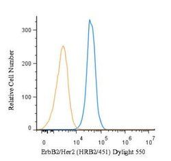
Antigen: ErbB2/Her2
Classification: Monoclonal
Conjugate: Unconjugated
Gene Alias: CD340, CD340 antigen, c-erb B2/neu protein, EC 2.7.10, EGFR2, HER-2, HER2EC 2.7.10.1, herstatin, Metastatic lymph node gene 19 protein, MLN 19, MLN19, NEUHER-2/neu, neuroblastoma/glioblastoma derived oncogene homolog, NGLTKR1, p185erbB2, Proto-oncogene c-ErbB-2, Proto-oncogene Neu, receptor tyrosine-protein kinase erbB-2, Tyrosine kinase-type cell surface receptor HER2, v-erb-b2 avian erythroblastic leukemia viral oncogene homolog 2(neuro/glioblastoma derived oncogene homolog), v-erb-b2 erythroblastic leukemia viral oncogene homolog 2, neuro/glioblastomaderived oncogene homolog (avian)
Host Species: Mouse
Purification Method: Protein A or G purified
Regulatory Status: RUO
Primary or Secondary: Primary
Test Specificity: This monoclonal antibody is specific to c-erbB-2/HER-2 and shows minimal cross-reaction with other members of the family. C-erbB-2/HER-2 is a member of the EGFR family. Receptors of this family are located on the plasma membrane and consist of an extracellular ligand-binding domain that is connected to a large intracellular domain by a single transmembrane sequence. c-erbB-2/HER-2 protein is over-expressed in a variety of carcinomas especially those of breast and ovary.
Content And Storage: Store at 4C short term. Aliquot and store at -20C long term. Avoid freeze-thaw cycles.
Isotype: IgG1 κ
Applications: Western Blot, Flow Cytometry, ELISA, Immunohistochemistry, Immunocytochemistry, Immunofluorescence, Immunoprecipitation, Immunohistochemistry (Paraffin), Immunohistochemistry (Frozen), Peptide Array, CyTOF
Clone: HRB2/451
Dilution: Western Blot 0.5-1ug/ml, Flow Cytometry 0.5-1ug/million cells, ELISA 1-5ug/ml for coating, Immunohistochemistry, Immunocytochemistry/Immunofluorescence 0.5-1ug/ml, Immunoprecipitation 0.5-1ug/500ug protein lysate, Immunohistochemistry-Paraffin, Immunohistochemistry-Frozen 0.5-1ug/ml, Protein Array, Flow (Cell Surface), CyTOF-ready
Gene Symbols: ERBB2
Immunogen: Recombinant human ErbB2/Her2 protein (Uniprot: IDP04626)
Quantity: 0.1 mg
Research Discipline: Breast Cancer, Cancer, Cellular Markers, Core ESC Like Genes, Phospho Specific, Protein Kinase, Stem Cell Markers, Tumor Suppressors
Gene ID (Entrez): 2064
Target Species: Human
Form: Purified