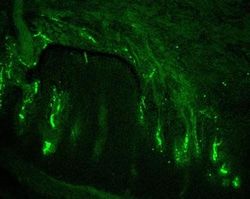
NF-H Antibody (SPM203) - IHC-Prediluted, Novus Biologicals™
Manufacturer: Novus Biologicals
Select a Size
| Pack Size | SKU | Availability | Price |
|---|---|---|---|
| Each of 1 | NBP248236-Each-of-1 | In Stock | ₹ 46,636.00 |
NBP248236 - Each of 1
In Stock
Quantity
1
Base Price: ₹ 46,636.00
GST (18%): ₹ 8,394.48
Total Price: ₹ 55,030.48
Antigen
NF-H
Classification
Monoclonal
Conjugate
Unconjugated
Formulation
10mM PBS and 0.05% BSA with 0.05% Sodium Azide
Gene Symbols
NEFH
Immunogen
Triton-X 100 insoluble protein fraction of rat brain (Uniprot: P12036)
Purification Method
Protein A or G purified
Regulatory Status
RUO
Primary or Secondary
Primary
Test Specificity
This monoclonal antibody reacts with a 200kDa protein, identified as heavy sub-unit of neurofilaments (NF-H). Neurofilaments make up the main structural elements of axons and dendrites and are found in neurons, peripheral nerves, and sympathetic ganglion cells. Neurofilaments consist of three major subunits with molecular weights of 68kDa (NF-L), 160kDa (NF-M) and 200kDa (NF-H). Anti-neurofilament stains a number of neural, neuroendocrine, and endocrine tumors. Neuromas, ganglioneuromas, gangliogliomas, ganglioneuroblastomas, and neuroblastomas stain positively for anti-neurofilament. Neurofilaments are also present in paragangliomas as well as adrenal and extra-adrenal pheochromocytomas. Carcinoids, neuroendocrine carcinomas of the skin, and oat cell carcinomas of the lung also express neurofilament.
Content And Storage
Store at 4C.
Isotype
IgG1 κ
Applications
Immunohistochemistry (Paraffin)
Clone
SPM203
Dilution
Immunohistochemistry-Paraffin
Gene Alias
200 kDa neurofilament protein, KIAA0845, Neurofilament Heavy (200kDa), neurofilament heavy polypeptide, Neurofilament triplet H protein, neurofilament, heavy polypeptide, neurofilament, heavy polypeptide 200kDa, NF200, NFH, NF-H
Host Species
Mouse
Molecular Weight of Antigen
200 kDa
Quantity
7 mL
Research Discipline
Autophagy, Cellular Markers, Cytoskeleton Markers, Neurodegeneration, Neurofilaments, Neuronal Cell Markers, Neuroscience
Gene ID (Entrez)
4744
Target Species
Human, Mouse, Rat, Porcine, Chicken
Form
Purified
Related Products


Description
- NF-H Monoclonal specifically detects NF-H in Human, Mouse, Rat, Porcine, Chicken samples
- It is validated for Immunohistochemistry, Immunohistochemistry-Paraffin.
Compare Similar Items
Show Difference
Antigen: NF-H
Classification: Monoclonal
Conjugate: Unconjugated
Formulation: 10mM PBS and 0.05% BSA with 0.05% Sodium Azide
Gene Symbols: NEFH
Immunogen: Triton-X 100 insoluble protein fraction of rat brain (Uniprot: P12036)
Purification Method: Protein A or G purified
Regulatory Status: RUO
Primary or Secondary: Primary
Test Specificity: This monoclonal antibody reacts with a 200kDa protein, identified as heavy sub-unit of neurofilaments (NF-H). Neurofilaments make up the main structural elements of axons and dendrites and are found in neurons, peripheral nerves, and sympathetic ganglion cells. Neurofilaments consist of three major subunits with molecular weights of 68kDa (NF-L), 160kDa (NF-M) and 200kDa (NF-H). Anti-neurofilament stains a number of neural, neuroendocrine, and endocrine tumors. Neuromas, ganglioneuromas, gangliogliomas, ganglioneuroblastomas, and neuroblastomas stain positively for anti-neurofilament. Neurofilaments are also present in paragangliomas as well as adrenal and extra-adrenal pheochromocytomas. Carcinoids, neuroendocrine carcinomas of the skin, and oat cell carcinomas of the lung also express neurofilament.
Content And Storage: Store at 4C.
Isotype: IgG1 κ
Applications: Immunohistochemistry (Paraffin)
Clone: SPM203
Dilution: Immunohistochemistry-Paraffin
Gene Alias: 200 kDa neurofilament protein, KIAA0845, Neurofilament Heavy (200kDa), neurofilament heavy polypeptide, Neurofilament triplet H protein, neurofilament, heavy polypeptide, neurofilament, heavy polypeptide 200kDa, NF200, NFH, NF-H
Host Species: Mouse
Molecular Weight of Antigen: 200 kDa
Quantity: 7 mL
Research Discipline: Autophagy, Cellular Markers, Cytoskeleton Markers, Neurodegeneration, Neurofilaments, Neuronal Cell Markers, Neuroscience
Gene ID (Entrez): 4744
Target Species: Human, Mouse, Rat, Porcine, Chicken
Form: Purified
Antigen: IgM Heavy Chain
Classification: Monoclonal
Conjugate: Unconjugated
Formulation: 10mM PBS and 0.05% BSA with 0.05% Sodium Azide
Gene Symbols: IGHM
Immunogen: Recombinant heavy chain of human IgM (IM260 & IM373); Heavy chain of human IgM (ICO-30)
Purification Method: Protein A or G purified
Regulatory Status: RUO
Primary or Secondary: Primary
Test Specificity: Recognizes a protein of 75kDa, identified as mu heavy chain of human immunoglobulins. It does not cross-react with alpha (IgA), gamma (IgG), epsilon (IgE), or delta (IgD), heavy chains, T-cells, monocytes, granulocytes, or erythrocytes. This monoclonal antibody is useful in the identification of leukemias, plasmacytomas, and certain non-Hodgkins lymphomas. The most common feature of these malignancies is the restricted expression of a single heavy chain class. Demonstration of clonality in lymphoid infiltrates indicates that the infiltrate is clonal and therefore malignant.
Content And Storage: Store at 4C.
Isotype: IgG1 κ, IgG2b κ
Applications: Immunohistochemistry (Paraffin)
Clone: Cocktail
Dilution: Immunohistochemistry-Paraffin
Gene Alias: Constant region of heavy chain of IgM, Ig Mu Chain C Region BOT Ig Mu Chain C Region GAL Ig Mu Chain C Region OU immunoglobulin heavy constant mu, Immunoglobulin mu chain
Host Species: Mouse
Molecular Weight of Antigen: __
Quantity: 7 mL
Research Discipline: __
Gene ID (Entrez): 3507
Target Species: Human
Form: Purified
Antigen: Insulin
Classification: Monoclonal
Conjugate: Unconjugated
Formulation: 10mM PBS and 0.05% BSA with 0.05% Sodium Azide
Gene Symbols: INS
Immunogen: Recombinant Insulin protein (Uniprot: P01308)
Purification Method: Protein A or G purified
Regulatory Status: RUO
Primary or Secondary: Primary
Test Specificity: Recognizes a polypeptide which is identified as insulin, a 51-amino acid polypeptide composed of A and B chains connected through the C-peptide. Proinsulin, which has very little biological activity, is cleaved by proteases within its cell of origin into the insulin molecule and the C-terminal basic residue. Insulin enhances membrane transport of glucose, amino acids, and certain ions. It also promotes glycogen storage, formation of triglycerides, and synthesis of proteins and nucleic acids. Deficiency of insulin results in diabetes mellitus. The main storage site for insulin is the pancreatic islets. Antibodies to insulin are important as beta-cell and insulinoma marker.
Content And Storage: Store at 4C.
Isotype: IgG1 κ
Applications: Immunohistochemistry (Paraffin)
Clone: IRDN/794
Dilution: Immunohistochemistry-Paraffin
Gene Alias: IDDM2, ILPR, insulin, IRDN, MODY10, proinsulin
Host Species: Mouse
Molecular Weight of Antigen: 6 kDa
Quantity: 7 mL
Research Discipline: Diabetes Research, Immune System Diseases, Immunology, Stem Cell Markers
Gene ID (Entrez): 3630
Target Species: Human, Mouse, Rat, Porcine, Primate
Form: Purified