NBP233096W

Thyroglobulin Antibody (6E1), Alexa Fluor™ 405, Novus Biologicals™

Manufacturer: Novus Biologicals

Select a Size

Pack Size SKU Availability Price
Each of 1 NBP233096W-Each-of-1 In Stock ₹ 55,271.76

NBP233096W - Each of 1

₹ 55,271.76

In Stock

Quantity

1

Base Price: ₹ 55,271.76

GST (18%): ₹ 9,948.917

Total Price: ₹ 65,220.677

Antigen

Thyroglobulin

Classification

Monoclonal

Conjugate

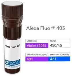
Alexa Fluor 405

Gene Alias

AITD3TGN, TDH3, Tg, thyroglobulin

Host Species

Mouse

Molecular Weight of Antigen

660 kDa

Quantity

0.1 mL

Research Discipline

Cancer, Cell Biology

Gene ID (Entrez)

7038

Target Species

Human, Mouse, Rat

Form

Purified

Applications

Western Blot, Flow Cytometry, Immunohistochemistry (Paraffin)

Clone

60

Dilution

Western Blot, Flow Cytometry, Immunohistochemistry-Paraffin

Gene Symbols

TG

Immunogen

Thyroglobulin isolated from human thyroid glands was used as the immunogen for the antibody.

Purification Method

Protein A or G purified

Regulatory Status

RUO

Primary or Secondary

Primary

Test Specificity

Thyroglobulin is a 660kDa dimeric pre-protein with multiple glycosylation sites. It is produced by and processed within the thyroid gland to produce the hormone thyroxine and triiodothyronine. Prior to forming dimers, thyroglobulin monomers undergo conformational maturation in the endoplasmic reticulation. The vast majority of follicular carcinomas of the thyroid will give positive immunoreactivity for anti-thyroglobulin even though sometimes only focally. Poorly differentiated carcinomas of the thyroid are frequently anti-thyroglobulin negative. Adenocarcinomas of other-than-thyroid origin do not react with this antibody. This antibody is useful in identification of thyroid carcinoma of the papillary and follicular types. Presence of thyroglobulin in metastatic lesions establishes the thyroid origin of tumor. Anti-thyroglobulin, combined with anti-calcitonin, can identify medullary carcinomas of the thyroid. Furthermore, anti-thyroglobulin, combined with anti-TTF1, can be a reliable m

Content And Storage

Store at 4C in the dark.

Isotype

IgG1 κ

Related Products

Img

Novus Biologicals

NBP233096G

--

Img

Novus Biologicals

NBP234441X

--

Img

Novus Biologicals

NBP248103

--

Img

Novus Biologicals

NB247773405

--

Img

Novus Biologicals

NB247773488

--

Img

Novus Biologicals

NBP248119

--

Img

Novus Biologicals

NB247773PCP

--

Img

Novus Biologicals

NB009163

--

Description

  • Thyroglobulin Monoclonal specifically detects Thyroglobulin in Human samples
  • It is validated for Immunohistochemistry, Immunohistochemistry-Paraffin.

Compare Similar Items

Show Difference

Img

Novus Biologicals

NBP233096W

--


Antigen:
Thyroglobulin

Classification:
Monoclonal

Conjugate:
Alexa Fluor 405

Gene Alias:
AITD3TGN, TDH3, Tg, thyroglobulin

Host Species:
Mouse

Molecular Weight of Antigen:
660 kDa

Quantity:
0.1 mL

Research Discipline:
Cancer, Cell Biology

Gene ID (Entrez):
7038

Target Species:
Human, Mouse, Rat

Form:
Purified

Applications:
Western Blot, Flow Cytometry, Immunohistochemistry (Paraffin)

Clone:
60

Dilution:
Western Blot, Flow Cytometry, Immunohistochemistry-Paraffin

Gene Symbols:
TG

Immunogen:
Thyroglobulin isolated from human thyroid glands was used as the immunogen for the antibody.

Purification Method:
Protein A or G purified

Regulatory Status:
RUO

Primary or Secondary:
Primary

Test Specificity:
Thyroglobulin is a 660kDa dimeric pre-protein with multiple glycosylation sites. It is produced by and processed within the thyroid gland to produce the hormone thyroxine and triiodothyronine. Prior to forming dimers, thyroglobulin monomers undergo conformational maturation in the endoplasmic reticulation. The vast majority of follicular carcinomas of the thyroid will give positive immunoreactivity for anti-thyroglobulin even though sometimes only focally. Poorly differentiated carcinomas of the thyroid are frequently anti-thyroglobulin negative. Adenocarcinomas of other-than-thyroid origin do not react with this antibody. This antibody is useful in identification of thyroid carcinoma of the papillary and follicular types. Presence of thyroglobulin in metastatic lesions establishes the thyroid origin of tumor. Anti-thyroglobulin, combined with anti-calcitonin, can identify medullary carcinomas of the thyroid. Furthermore, anti-thyroglobulin, combined with anti-TTF1, can be a reliable m

Content And Storage:
Store at 4C in the dark.

Isotype:
IgG1 κ

Img

Novus Biologicals

NBP233096Y

--


Antigen:
Thyroglobulin

Classification:
Monoclonal

Conjugate:
Alexa Fluor 647

Gene Alias:
AITD3TGN, TDH3, Tg, thyroglobulin

Host Species:
Mouse

Molecular Weight of Antigen:
660 kDa

Quantity:
0.1 mL

Research Discipline:
Cancer, Cell Biology

Gene ID (Entrez):
7038

Target Species:
Human, Mouse, Rat

Form:
Purified

Applications:
Western Blot, Flow Cytometry, Immunohistochemistry (Paraffin)

Clone:
60

Dilution:
Western Blot, Flow Cytometry, Immunohistochemistry-Paraffin

Gene Symbols:
TG

Immunogen:
Thyroglobulin isolated from human thyroid glands was used as the immunogen for the antibody.

Purification Method:
Protein A or G purified

Regulatory Status:
RUO

Primary or Secondary:
Primary

Test Specificity:
Thyroglobulin is a 660kDa dimeric pre-protein with multiple glycosylation sites. It is produced by and processed within the thyroid gland to produce the hormone thyroxine and triiodothyronine. Prior to forming dimers, thyroglobulin monomers undergo conformational maturation in the endoplasmic reticulation. The vast majority of follicular carcinomas of the thyroid will give positive immunoreactivity for anti-thyroglobulin even though sometimes only focally. Poorly differentiated carcinomas of the thyroid are frequently anti-thyroglobulin negative. Adenocarcinomas of other-than-thyroid origin do not react with this antibody. This antibody is useful in identification of thyroid carcinoma of the papillary and follicular types. Presence of thyroglobulin in metastatic lesions establishes the thyroid origin of tumor. Anti-thyroglobulin, combined with anti-calcitonin, can identify medullary carcinomas of the thyroid. Furthermore, anti-thyroglobulin, combined with anti-TTF1, can be a reliable m

Content And Storage:
Store at 4C in the dark.

Isotype:
IgG1 κ

Img

Novus Biologicals

NBP233098

--


Antigen:
TIMP-3

Classification:
Polyclonal

Conjugate:
Unconjugated

Gene Alias:
HSMRK222, K222, K222TA2, metalloproteinase inhibitor 3, MIG-5 protein, Protein MIG-5, pseudoinflammatory), SFD, TIMP metallopeptidase inhibitor 3, TIMP-3, tissue inhibitor of metalloproteinase 3 (Sorsby fundus dystrophy, Tissue inhibitor of metalloproteinases 3

Host Species:
Rabbit

Molecular Weight of Antigen:
__

Quantity:
0.1 mg

Research Discipline:
Angiogenesis, Apoptosis, Cancer, Hypoxia, Vision

Gene ID (Entrez):
7078

Target Species:
Human, Bovine, Canine, Equine, Sheep

Form:
Purified

Applications:
Western Blot, Flow Cytometry, Immunohistochemistry, Immunocytochemistry, Immunofluorescence, Immunoprecipitation

Clone:
__

Dilution:
Western Blot, Flow Cytometry 0.5-1ug/million cells, Immunohistochemistry 0.5-1ug/ml, Immunocytochemistry/Immunofluorescence 1-2ug/ml, Immunoprecipitation 0.5-1ug/500ug protein lysate, Immunohistochemistry-Paraffin 0.5-1ug/ml, CyTOF-ready

Gene Symbols:
TIMP3

Immunogen:
TIMP-3 (aa175-211) (Uniprot: P35625)

Purification Method:
Protein A purified

Regulatory Status:
RUO

Primary or Secondary:
Primary

Test Specificity:
TIMP3 (tissue inhibitor of metalloproteinases 3), along with family members TIMP1, TIMP2, and TIMP4, are inhibitors of the matrix metalloproteinases (MMPs), a group of peptidases involved in degradation of the extracellular matrix (ECM). An imbalance between MMPs and the associated TIMPs may play a significant role in the invasive phenotype of malignant tumors. TIMP s inhibit the proteolytic invasiveness of tumor cells and normal placental trophoblast cells. TIMP-3 may be involved in regulating trophoblastic invasion of the uterus as well as in regulating remodeling of the extracellular matrix during the folding of epithelia, and in the formation, branching and expansion of epithelial tubes.

Content And Storage:
Store at 4C short term. Aliquot and store at -20C long term. Avoid freeze-thaw cycles.

Isotype:
IgG

Img

Novus Biologicals

NBP233102

--


Antigen:
NF-H

Classification:
Monoclonal

Conjugate:
Unconjugated

Gene Alias:
200 kDa neurofilament protein, KIAA0845, Neurofilament Heavy (200kDa), neurofilament heavy polypeptide, Neurofilament triplet H protein, neurofilament, heavy polypeptide, neurofilament, heavy polypeptide 200kDa, NF200, NFH, NF-H

Host Species:
Mouse

Molecular Weight of Antigen:
200 kDa

Quantity:
0.1 mg

Research Discipline:
Autophagy, Cellular Markers, Cytoskeleton Markers, Neurodegeneration, Neurofilaments, Neuronal Cell Markers, Neuroscience

Gene ID (Entrez):
4744

Target Species:
Human, Mouse, Rat, Porcine, Chicken

Form:
Purified

Applications:
ELISA

Clone:
NF421

Dilution:
ELISA 1-5ug/ml for coating

Gene Symbols:
NEFH

Immunogen:
Recombinant full-length human NF-H protein (Uniprot: P12036)

Purification Method:
Protein A or G purified

Regulatory Status:
RUO

Primary or Secondary:
Primary

Test Specificity:
This monoclonal antibody reacts with a 200kDa protein, identified as heavy sub-unit of neurofilaments (NF-H). Neurofilaments make up the main structural elements of axons and dendrites and are found in neurons, peripheral nerves, and sympathetic ganglion cells. Neurofilaments consist of three major subunits with molecular weights of 68kDa (NF-L), 160kDa (NF-M) and 200kDa (NF-H). Anti-neurofilament stains a number of neural, neuroendocrine, and endocrine tumors. Neuromas, ganglioneuromas, gangliogliomas, ganglioneuroblastomas, and neuroblastomas stain positively for anti-neurofilament. Neurofilaments are also present in paragangliomas as well as adrenal and extra-adrenal pheochromocytomas. Carcinoids, neuroendocrine carcinomas of the skin, and oat cell carcinomas of the lung also express neurofilament.

Content And Storage:
Store at 4C short term. Aliquot and store at -20C long term. Avoid freeze-thaw cycles.

Isotype:
IgG1 κ