CD31/PECAM-1 Antibody (C31.7), Alexa Fluor™ 647, Novus Biologicals™
Manufacturer: Novus Biologicals
Select a Size
| Pack Size | SKU | Availability | Price |
|---|---|---|---|
| Each of 1 | NBP233136Y-Each-of-1 | In Stock | ₹ 57,494.00 |
NBP233136Y - Each of 1
In Stock
Quantity
1
Base Price: ₹ 57,494.00
GST (18%): ₹ 10,348.92
Total Price: ₹ 67,842.92
Antigen
CD31/PECAM-1
Classification
Monoclonal
Conjugate
Alexa Fluor 647
Gene Alias
adhesion molecule, CD31, CD31 antigen, CD31/EndoCAM, EndoCAM, FLJ34100, FLJ58394, GPIIA′, PECA1, PECAM-1, PECAM-1, CD31/EndoCAM, platelet endothelial cell adhesion molecule, platelet endothelial cell adhesion molecule-1, platelet/endothelial cell adhesion molecule
Host Species
Mouse
Molecular Weight of Antigen
82.5 kDa
Quantity
0.1 mL
Research Discipline
Angiogenesis, Cancer, Cellular Markers, Cytoskeleton Markers, Embryonic Stem Cell Markers, Endothelial Cell Markers, Extracellular Matrix, Hematopoietic Stem Cell Markers, Immunology, Mesenchymal Stem Cell Markers, Myeloid Cell Markers, Signal Transduction, Stem Cell Markers
Gene ID (Entrez)
5175
Target Species
Human, Cynomolgus Monkey, Rabbit
Form
Purified
Applications
Western Blot, Flow Cytometry, Immunocytochemistry, Immunofluorescence, Immunohistochemistry (Paraffin), Immunohistochemistry (Frozen)
Clone
C31.7
Dilution
Western Blot, Flow Cytometry, Immunocytochemistry/Immunofluorescence, Immunohistochemistry-Paraffin, Immunohistochemistry-Frozen
Gene Symbols
PECAM1
Immunogen
Recombinant human full-length CD31/PECAM-1 protein (Uniprot: P16284)
Purification Method
Protein A or G purified
Regulatory Status
RUO
Primary or Secondary
Primary
Test Specificity
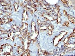
CD31 (PECAM-1) is a transmembrane glycoprotein member of the immunoglobulin supergene family of adhesion molecules. CD31 is expressed by stem cells of the hematopoietic system and is primarily used to identify and concentrate these cells for experimental studies as well as for bone marrow transplantation. Anti-CD31 has shown to be highly specific and sensitive for vascular endothelial cells. Staining of nonvascular tumors (excluding hematopoietic neoplasms) is rare. CD31 monoclonal antibody reacts with normal, benign, and malignant endothelial cells which make up blood vessel lining. The level of CD31 expression can help to determine the degree of tumor angiogenesis, and a high level of CD31 expression may imply a rapidly growing tumor and potentially a predictor of tumor recurrence.
Content And Storage
Store at 4C in the dark.
Isotype
IgG1 κ
Related Products

Description
- CD31/PECAM-1 Monoclonal specifically detects CD31/PECAM-1 in Human, Cynomolgus Monkey, Rabbit samples
- It is validated for Western Blot, Flow Cytometry, Immunohistochemistry, Immunohistochemistry-Paraffin.
Compare Similar Items
Show Difference
Antigen: CD31/PECAM-1
Classification: Monoclonal
Conjugate: Alexa Fluor 647
Gene Alias: adhesion molecule, CD31, CD31 antigen, CD31/EndoCAM, EndoCAM, FLJ34100, FLJ58394, GPIIA′, PECA1, PECAM-1, PECAM-1, CD31/EndoCAM, platelet endothelial cell adhesion molecule, platelet endothelial cell adhesion molecule-1, platelet/endothelial cell adhesion molecule
Host Species: Mouse
Molecular Weight of Antigen: 82.5 kDa
Quantity: 0.1 mL
Research Discipline: Angiogenesis, Cancer, Cellular Markers, Cytoskeleton Markers, Embryonic Stem Cell Markers, Endothelial Cell Markers, Extracellular Matrix, Hematopoietic Stem Cell Markers, Immunology, Mesenchymal Stem Cell Markers, Myeloid Cell Markers, Signal Transduction, Stem Cell Markers
Gene ID (Entrez): 5175
Target Species: Human, Cynomolgus Monkey, Rabbit
Form: Purified
Applications: Western Blot, Flow Cytometry, Immunocytochemistry, Immunofluorescence, Immunohistochemistry (Paraffin), Immunohistochemistry (Frozen)
Clone: C31.7
Dilution: Western Blot, Flow Cytometry, Immunocytochemistry/Immunofluorescence, Immunohistochemistry-Paraffin, Immunohistochemistry-Frozen
Gene Symbols: PECAM1
Immunogen: Recombinant human full-length CD31/PECAM-1 protein (Uniprot: P16284)
Purification Method: Protein A or G purified
Regulatory Status: RUO
Primary or Secondary: Primary
Test Specificity: CD31 (PECAM-1) is a transmembrane glycoprotein member of the immunoglobulin supergene family of adhesion molecules. CD31 is expressed by stem cells of the hematopoietic system and is primarily used to identify and concentrate these cells for experimental studies as well as for bone marrow transplantation. Anti-CD31 has shown to be highly specific and sensitive for vascular endothelial cells. Staining of nonvascular tumors (excluding hematopoietic neoplasms) is rare. CD31 monoclonal antibody reacts with normal, benign, and malignant endothelial cells which make up blood vessel lining. The level of CD31 expression can help to determine the degree of tumor angiogenesis, and a high level of CD31 expression may imply a rapidly growing tumor and potentially a predictor of tumor recurrence.
Content And Storage: Store at 4C in the dark.
Isotype: IgG1 κ
Antigen: CD31/PECAM-1
Classification: Monoclonal
Conjugate: Alexa Fluor 700
Gene Alias: adhesion molecule, CD31, CD31 antigen, CD31/EndoCAM, EndoCAM, FLJ34100, FLJ58394, GPIIA′, PECA1, PECAM-1, PECAM-1, CD31/EndoCAM, platelet endothelial cell adhesion molecule, platelet endothelial cell adhesion molecule-1, platelet/endothelial cell adhesion molecule
Host Species: Mouse
Molecular Weight of Antigen: 82.5 kDa
Quantity: 0.1 mL
Research Discipline: Angiogenesis, Cancer, Cellular Markers, Cytoskeleton Markers, Embryonic Stem Cell Markers, Endothelial Cell Markers, Extracellular Matrix, Hematopoietic Stem Cell Markers, Immunology, Mesenchymal Stem Cell Markers, Myeloid Cell Markers, Signal Transduction, Stem Cell Markers
Gene ID (Entrez): 5175
Target Species: Human, Cynomolgus Monkey, Rabbit
Form: Purified
Applications: Western Blot, Flow Cytometry, Immunocytochemistry, Immunofluorescence, Immunohistochemistry (Paraffin), Immunohistochemistry (Frozen)
Clone: C31.7
Dilution: Western Blot, Flow Cytometry, Immunocytochemistry/Immunofluorescence, Immunohistochemistry-Paraffin, Immunohistochemistry-Frozen
Gene Symbols: PECAM1
Immunogen: Recombinant human full-length CD31/PECAM-1 protein (Uniprot: P16284)
Purification Method: Protein A or G purified
Regulatory Status: RUO
Primary or Secondary: Primary
Test Specificity: CD31 (PECAM-1) is a transmembrane glycoprotein member of the immunoglobulin supergene family of adhesion molecules. CD31 is expressed by stem cells of the hematopoietic system and is primarily used to identify and concentrate these cells for experimental studies as well as for bone marrow transplantation. Anti-CD31 has shown to be highly specific and sensitive for vascular endothelial cells. Staining of nonvascular tumors (excluding hematopoietic neoplasms) is rare. CD31 monoclonal antibody reacts with normal, benign, and malignant endothelial cells which make up blood vessel lining. The level of CD31 expression can help to determine the degree of tumor angiogenesis, and a high level of CD31 expression may imply a rapidly growing tumor and potentially a predictor of tumor recurrence.
Content And Storage: Store at 4C in the dark.
Isotype: IgG1 κ

Antigen: Cyclin D1
Classification: Monoclonal
Conjugate: Unconjugated
Gene Alias: B-cell lymphoma 1 protein, BCL-1, BCL-1 oncogene, BCL1D11S287E, cyclin D1, cyclin D1 (PRAD1: parathyroid adenomatosis 1), G1/S-specific cyclin D1, G1/S-specific cyclin-D1, PRAD1 oncogene, PRAD1B-cell CLL/lymphoma 1, U21B31
Host Species: Mouse
Molecular Weight of Antigen: __
Quantity: 0.1 mg
Research Discipline: Cancer, Cell Cycle and Replication, Core ESC Like Genes, mTOR Pathway, Stem Cell Markers, Wnt Signaling Pathway
Gene ID (Entrez): 595
Target Species: Human, Mouse, Rat, Primate
Form: Purified
Applications: Western Blot, Flow Cytometry, ELISA, Immunohistochemistry (Paraffin), CyTOF
Clone: DCS-6
Dilution: Western Blot, Flow Cytometry, ELISA 1-5ug/ml for coating, Immunohistochemistry-Paraffin, CyTOF-ready
Gene Symbols: CCND1
Immunogen: Human full length recombinant cyclin D1 protein (Uniprot: P24385)
Purification Method: Protein A or G purified
Regulatory Status: RUO
Primary or Secondary: Primary
Test Specificity: Recognizes a protein of 36kDa, identified as cyclin D1. Cyclin D1, one of the key cell cycle regulators, is a putative proto-oncogene overexpressed in a wide variety of human neoplasms. This antibody neutralizes the activity of cyclin D1 in vivo. About 60% of mantle cell lymphomas (MCL) contain a t(11; 14)(q13; q32) translocation resulting in over-expression of cyclin D1. This antibody is useful in identifying mantle cell lymphomas (cyclin D1 positive) from CLL/SLL and follicular lymphomas (cyclin D1 negative). About 40% of breast carcinomas are positive for Cyclin D1. Occasionally, hairy cell leukemia and plasma cell myeloma weakly express Cyclin D1.
Content And Storage: Store at 4C short term. Aliquot and store at -20C long term. Avoid freeze-thaw cycles.
Isotype: IgG2a κ