EBV Early Antigens Antibody (1108-1), Alexa Fluor™ 405, Novus Biologicals™
Manufacturer: Novus Biologicals
Select a Size
| Pack Size | SKU | Availability | Price |
|---|---|---|---|
| Each of 1 | NBP234516W-Each-of-1 | In Stock | ₹ 57,494.00 |
NBP234516W - Each of 1
In Stock
Quantity
1
Base Price: ₹ 57,494.00
GST (18%): ₹ 10,348.92
Total Price: ₹ 67,842.92
Antigen
EBV Early Antigens
Classification
Monoclonal
Conjugate
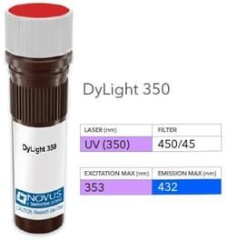
Alexa Fluor 405
Gene Alias
Chronic ulcerative stomatitis protein, CUSP, Keratinocyte transcription factor KET, p40, p51, p63, p73L, TP63, Transformation-related protein 63, Tumor protein 63, Tumor protein p73-like
Immunogen
Affinity purified early antigen polypeptides from induced Raji cells precipitated with African Burkitt's lymphoma sera
Quantity
0.1 mL
Primary or Secondary
Primary
Target Species
Virus
Form
Purified
Applications
Western Blot, Immunocytochemistry, Immunofluorescence, Immunohistochemistry (Frozen)
Clone
1108-1
Dilution
Western Blot, Immunocytochemistry/Immunofluorescence, Immunohistochemistry-Frozen
Host Species
Mouse
Purification Method
Protein A or G purified
Regulatory Status
RUO
Test Specificity
Epstein-Barr virus (EBV), also designated human herpesvirus 4 (HHV-4), is a member of the herpesvirus family and is one of the most common human viruses. EBV infects B cells and, though often asymptomatic, it can cause infectious mononucleosis, a disease characterized by fatigue, fever, sore throat and muscle soreness. The EBV-induced early antigens (Ea) are among several antigen complexes that have been identified in EBV-infected cells. The Ea complex is composed of diffuse (Ea-D) and restricted (Ea-R) components. The activity of Ea-D is suppressed during latent infection. BMRF1, the gene that encodes for Ea-D, is closely associated with the gene encoding for EBV DNA polymerase, and Ea-D is essential for the activity of this polymerase. Ea-D forms a complex with EBV DNase and, together, they may play a role in viral replication.
Content And Storage
Store at 4C in the dark.
Isotype
IgG1 κ
Related Products

Description
- EBV Early Antigens Monoclonal specifically detects EBV Early Antigens in Virus samples
- It is validated for Immunocytochemistry/Immunofluorescence.
Compare Similar Items
Show Difference
Antigen: EBV Early Antigens
Classification: Monoclonal
Conjugate: Alexa Fluor 405
Gene Alias: Chronic ulcerative stomatitis protein, CUSP, Keratinocyte transcription factor KET, p40, p51, p63, p73L, TP63, Transformation-related protein 63, Tumor protein 63, Tumor protein p73-like
Immunogen: Affinity purified early antigen polypeptides from induced Raji cells precipitated with African Burkitt's lymphoma sera
Quantity: 0.1 mL
Primary or Secondary: Primary
Target Species: Virus
Form: Purified
Applications: Western Blot, Immunocytochemistry, Immunofluorescence, Immunohistochemistry (Frozen)
Clone: 1108-1
Dilution: Western Blot, Immunocytochemistry/Immunofluorescence, Immunohistochemistry-Frozen
Host Species: Mouse
Purification Method: Protein A or G purified
Regulatory Status: RUO
Test Specificity: Epstein-Barr virus (EBV), also designated human herpesvirus 4 (HHV-4), is a member of the herpesvirus family and is one of the most common human viruses. EBV infects B cells and, though often asymptomatic, it can cause infectious mononucleosis, a disease characterized by fatigue, fever, sore throat and muscle soreness. The EBV-induced early antigens (Ea) are among several antigen complexes that have been identified in EBV-infected cells. The Ea complex is composed of diffuse (Ea-D) and restricted (Ea-R) components. The activity of Ea-D is suppressed during latent infection. BMRF1, the gene that encodes for Ea-D, is closely associated with the gene encoding for EBV DNA polymerase, and Ea-D is essential for the activity of this polymerase. Ea-D forms a complex with EBV DNase and, together, they may play a role in viral replication.
Content And Storage: Store at 4C in the dark.
Isotype: IgG1 κ
Antigen: EBV Early Antigens
Classification: Monoclonal
Conjugate: Alexa Fluor 488
Gene Alias: Chronic ulcerative stomatitis protein, CUSP, Keratinocyte transcription factor KET, p40, p51, p63, p73L, TP63, Transformation-related protein 63, Tumor protein 63, Tumor protein p73-like
Immunogen: Affinity purified early antigen polypeptides from induced Raji cells precipitated with African Burkitt's lymphoma sera
Quantity: 0.1 mL
Primary or Secondary: Primary
Target Species: Virus
Form: Purified
Applications: Western Blot, Immunocytochemistry, Immunofluorescence, Immunohistochemistry (Frozen)
Clone: 1108-1
Dilution: Western Blot, Immunocytochemistry/Immunofluorescence, Immunohistochemistry-Frozen
Host Species: Mouse
Purification Method: Protein A or G purified
Regulatory Status: RUO
Test Specificity: Epstein-Barr virus (EBV), also designated human herpesvirus 4 (HHV-4), is a member of the herpesvirus family and is one of the most common human viruses. EBV infects B cells and, though often asymptomatic, it can cause infectious mononucleosis, a disease characterized by fatigue, fever, sore throat and muscle soreness. The EBV-induced early antigens (Ea) are among several antigen complexes that have been identified in EBV-infected cells. The Ea complex is composed of diffuse (Ea-D) and restricted (Ea-R) components. The activity of Ea-D is suppressed during latent infection. BMRF1, the gene that encodes for Ea-D, is closely associated with the gene encoding for EBV DNA polymerase, and Ea-D is essential for the activity of this polymerase. Ea-D forms a complex with EBV DNase and, together, they may play a role in viral replication.
Content And Storage: Store at 4C in the dark.
Isotype: IgG1 κ
Antigen: Mitochondria
Classification: Monoclonal
Conjugate: Biotin
Gene Alias: __
Immunogen: Semi-purified mitochondrial preparation
Quantity: 0.1 mL
Primary or Secondary: Primary
Target Species: Human, Mouse (Negative), Rat (Negative)
Form: Purified
Applications: Western Blot, Immunocytochemistry, Immunofluorescence, Immunohistochemistry (Paraffin), Immunohistochemistry (Frozen)
Clone: 113-1
Dilution: Western Blot, Immunocytochemistry/Immunofluorescence, Immunohistochemistry-Paraffin, Immunohistochemistry-Frozen
Host Species: Mouse
Purification Method: Protein A or G purified
Regulatory Status: RUO
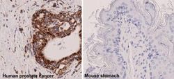
Test Specificity: This monoclonal antibody recognizes a 60kDa antigen associated with the mitochondria in human cells. It can be used to stain mitochondria in cell or tissue preparations and can be used as a mitochondrial marker in subcellular fractions. It produces a spaghetti-like pattern in normal and malignant cells. This antibody is an excellent marker for human cells in xenographic model research. It reacts specifically with human cells, including neurons and embryonic stem cells. Immunostaining pattern with anti-mitochondrial monoclonal antibody has been reported as a useful discriminatory adjunct in the complex differential diagnosis of granular renal cell tumors. Reportedly, this monoclonal antibody facilitates the classification of salivary tumors.
Content And Storage: Store at 4C in the dark.
Isotype: IgG1 κ