MVP Antibody (1032), Novus Biologicals™
Manufacturer: Fischer Scientific
The price for this product is unavailable. Please request a quote
Antigen
MVP
Classification
Monoclonal
Concentration
0.2mg/mL
Dilution
Flow Cytometry 0.5 - 1 ug/million cells in 0.1 ml, Immunohistochemistry-Paraffin 0.5 - 1.0 ug/ml, Immunofluorescence 0.5 - 1.0 ug/ml
Gene Accession No.
Q14764
Gene Symbols
MVP
Immunogen
Proteins precipitated from human breast cancer MCF-7 cells
Quantity
0.2 mg
Research Discipline
Cancer
Gene ID (Entrez)
9961
Target Species
Human
Form
Purified
Applications
Flow Cytometry, Immunohistochemistry (Paraffin), Immunofluorescence
Clone
1032
Conjugate
Unconjugated
Formulation
10mM PBS and 0.05% BSA with 0.05% Sodium Azide
Gene Alias
LRPVAULT1, Lung resistance-related protein, major vault protein
Host Species
Mouse
Purification Method
Protein A or G purified
Regulatory Status
RUO
Primary or Secondary
Primary
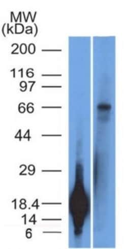
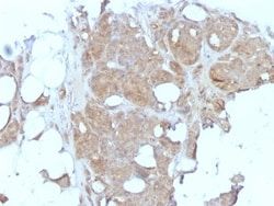
Test Specificity
Recognizes a protein of 104kDa-110kDa, characterized as major vault protein (MVP). Vaults are large ribonucleoprotein particles (RNPs) present in all eukaryotic cells. They have a complex morphology, including several small molecules of RNA, but a single protein species. The MVP accounts for >70% of their mass. Their shape is reminiscent of the nucleopore central plug. Treatment of cells with estradiol increases the amount of MVP in nuclear extract. The hormone-dependent interaction of vaults with ER is prevented in vitro by sodium molybdate. Antibodies to estrogen, progesterone and glucocorticoid receptors are able to co-immunoprecipitate the MVP. MVP is overexpressed in many neoplastic tissues and cell lines. Expression of MVP predicts a poor response to chemotherapy.
Content And Storage
Store at 4C.
Isotype
IgG1 κ
Description
- Ensure accurate, reproducible results in Flow Cytometry, Immunohistochemistry (Paraffin), Immunofluorescence MVP Monoclonal specifically detects MVP in Human samples
- It is validated for Flow Cytometry, Immunohistochemistry, Immunocytochemistry/Immunofluorescence, Immunohistochemistry-Paraffin, Immunofluorescence.
Compare Similar Items
Show Difference
Antigen: MVP
Classification: Monoclonal
Concentration: 0.2mg/mL
Dilution: Flow Cytometry 0.5 - 1 ug/million cells in 0.1 ml, Immunohistochemistry-Paraffin 0.5 - 1.0 ug/ml, Immunofluorescence 0.5 - 1.0 ug/ml
Gene Accession No.: Q14764
Gene Symbols: MVP
Immunogen: Proteins precipitated from human breast cancer MCF-7 cells
Quantity: 0.2 mg
Research Discipline: Cancer
Gene ID (Entrez): 9961
Target Species: Human
Form: Purified
Applications: Flow Cytometry, Immunohistochemistry (Paraffin), Immunofluorescence
Clone: 1032
Conjugate: Unconjugated
Formulation: 10mM PBS and 0.05% BSA with 0.05% Sodium Azide
Gene Alias: LRPVAULT1, Lung resistance-related protein, major vault protein
Host Species: Mouse
Purification Method: Protein A or G purified
Regulatory Status: RUO
Primary or Secondary: Primary
Test Specificity: Recognizes a protein of 104kDa-110kDa, characterized as major vault protein (MVP). Vaults are large ribonucleoprotein particles (RNPs) present in all eukaryotic cells. They have a complex morphology, including several small molecules of RNA, but a single protein species. The MVP accounts for >70% of their mass. Their shape is reminiscent of the nucleopore central plug. Treatment of cells with estradiol increases the amount of MVP in nuclear extract. The hormone-dependent interaction of vaults with ER is prevented in vitro by sodium molybdate. Antibodies to estrogen, progesterone and glucocorticoid receptors are able to co-immunoprecipitate the MVP. MVP is overexpressed in many neoplastic tissues and cell lines. Expression of MVP predicts a poor response to chemotherapy.
Content And Storage: Store at 4C.
Isotype: IgG1 κ

Antigen: TRIM29
Classification: Monoclonal
Concentration: 0.2mg/mL
Dilution: Western Blot 0.5 - 1.0 ug/ml, Flow Cytometry 0.5 - 1 ug/million cells in 0.1 ml, SDS-Page, Immunofluorescence 0.5 - 1.0 ug/ml
Gene Accession No.: Q14134
Gene Symbols: TRIM29
Immunogen: Recombinant fragment (126 Amino acid residues between aa 1-200) of human TRIM29 protein
Quantity: 0.02 mg
Research Discipline: __
Gene ID (Entrez): 23650
Target Species: Human
Form: Purified
Applications: Western Blot, Flow Cytometry, SDS-Page, Immunofluorescence
Clone: TRIM29/1042
Conjugate: Unconjugated
Formulation: 1.0mM PBS and 0.05% BSA with 0.05% Sodium Azide
Gene Alias: Ataxia telangiectasia group D-associated protein, ATDCataxia-telangiectasia group D-associated protein, FLJ36085, tripartite motif containing 29, tripartite motif protein TRIM29, tripartite motif-containing 29, tripartite motif-containing protein 29
Host Species: Mouse
Purification Method: Protein A or G purified
Regulatory Status: RUO
Primary or Secondary: Primary
Test Specificity: It recognizes a 66kDa protein, which is identified as Tripartite motif-containing protein 29 (TRIM29). It interacts with the intermediate filament protein vimentin, a substrate for the PKC family of protein kinases, and with hPKCI-1, an inhibitor of the PKCs. TRIM29 protein contains both zinc finger and leucine zipper motifs, suggesting that the it may form homodimers and possibly associate with DNA. High expression of TRIM29 has been reported in gastric cancer and pancreatic cancer, and correlates with enhanced tumor growth and lymph node metastasis. TRIM29 is also able to distinguish lung squamous cell carcinoma from lung adenocarcinoma with ∼90% positive accuracy, when used in a panel with TTF-1, p63, CK5/6, and Napsin-A antibodies.
Content And Storage: Store at 4C.
Isotype: IgG2b κ
Antigen: TRIM29
Classification: Monoclonal
Concentration: 0.2mg/mL
Dilution: Western Blot 0.5 - 1.0 ug/ml, Flow Cytometry 0.5 - 1 ug/million cells in 0.1 ml, SDS-Page, Immunofluorescence 0.5 - 1.0 ug/ml
Gene Accession No.: Q14134
Gene Symbols: TRIM29
Immunogen: Recombinant fragment (126 Amino acid residues between aa 1-200) of human TRIM29 protein
Quantity: 0.1 mg
Research Discipline: __
Gene ID (Entrez): 23650
Target Species: Human
Form: Purified
Applications: Western Blot, Flow Cytometry, SDS-Page, Immunofluorescence
Clone: TRIM29/1042
Conjugate: Unconjugated
Formulation: 1.0mM PBS and 0.05% BSA with 0.05% Sodium Azide
Gene Alias: Ataxia telangiectasia group D-associated protein, ATDCataxia-telangiectasia group D-associated protein, FLJ36085, tripartite motif containing 29, tripartite motif protein TRIM29, tripartite motif-containing 29, tripartite motif-containing protein 29
Host Species: Mouse
Purification Method: Protein A or G purified
Regulatory Status: RUO
Primary or Secondary: Primary
Test Specificity: It recognizes a 66kDa protein, which is identified as Tripartite motif-containing protein 29 (TRIM29). It interacts with the intermediate filament protein vimentin, a substrate for the PKC family of protein kinases, and with hPKCI-1, an inhibitor of the PKCs. TRIM29 protein contains both zinc finger and leucine zipper motifs, suggesting that the it may form homodimers and possibly associate with DNA. High expression of TRIM29 has been reported in gastric cancer and pancreatic cancer, and correlates with enhanced tumor growth and lymph node metastasis. TRIM29 is also able to distinguish lung squamous cell carcinoma from lung adenocarcinoma with ∼90% positive accuracy, when used in a panel with TTF-1, p63, CK5/6, and Napsin-A antibodies.
Content And Storage: Store at 4C.
Isotype: IgG2b κ