CEACAM5/CD66e Antibody (C66/1030), Novus Biologicals™
Manufacturer: Fischer Scientific
The price for this product is unavailable. Please request a quote
Antigen
CEACAM5/CD66e
Classification
Monoclonal
Concentration
0.2mg/mL
Dilution
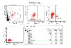
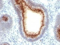
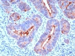
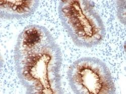
Flow Cytometry 0.5 - 1 ug/million cells in 0.1 ml, Immunohistochemistry-Paraffin 0.25 - 0.5 ug/ml, Immunofluorescence 1 - 2 ug/ml
Gene Accession No.
P06731
Gene Symbols
CEACAM5
Immunogen
Recombinant full-length human CEA protein
Quantity
0.2 mg
Research Discipline
Cancer, Cellular Markers, Immunology
Gene ID (Entrez)
1048
Target Species
Human
Form
Purified
Applications
Flow Cytometry, Immunohistochemistry (Paraffin), Immunofluorescence
Clone
C66/1030
Conjugate
Unconjugated
Formulation
10mM PBS and 0.05% BSA with 0.05% Sodium Azide
Gene Alias
Carcinoembryonic antigen, carcinoembryonic antigen-related cell adhesion molecule 5, CD66e antigen, CEACD66e, DKFZp781M2392, Meconium antigen 100
Host Species
Mouse
Purification Method
Protein A or G purified
Regulatory Status
RUO
Primary or Secondary
Primary
Test Specificity
This antibody recognizes proteins of 80-200kDa, identified as different members of CEA family. CEA is synthesized during development in the fetal gut and is re-expressed in increased amounts in intestinal carcinomas and several other tumors. This MAb does not react with nonspecific cross-reacting antigen (NCA) and with human polymorphonuclear leucocytes. It shows no reaction with a variety of normal tissues and is suitable for staining of formalin/paraffin tissues. CEA is not found in benign glands, stroma, or malignant prostatic cells. Antibody to CEA is useful in detecting early foci of gastric carcinoma and in distinguishing pulmonary adenocarcinomas (60-70% are CEA+) from pleural mesotheliomas (rarely or weakly CEA+). Anti-CEA positivity is seen in adenocarcinomas from the lung, colon, stomach, esophagus, pancreas, gallbadder, urachus, salivary gland, ovary, and endocervix
Content And Storage
Store at 4C.
Isotype
IgG1 κ
Related Products


Description
- Ensure accurate, reproducible results in Flow Cytometry, Immunohistochemistry (Paraffin), Immunofluorescence CEACAM5/CD66e Monoclonal specifically detects CEACAM5/CD66e in Human samples
- It is validated for Flow Cytometry, Immunohistochemistry, Immunocytochemistry/Immunofluorescence, Immunohistochemistry-Paraffin, Immunofluorescence.
Compare Similar Items
Show Difference
Antigen: CEACAM5/CD66e
Classification: Monoclonal
Concentration: 0.2mg/mL
Dilution: Flow Cytometry 0.5 - 1 ug/million cells in 0.1 ml, Immunohistochemistry-Paraffin 0.25 - 0.5 ug/ml, Immunofluorescence 1 - 2 ug/ml
Gene Accession No.: P06731
Gene Symbols: CEACAM5
Immunogen: Recombinant full-length human CEA protein
Quantity: 0.2 mg
Research Discipline: Cancer, Cellular Markers, Immunology
Gene ID (Entrez): 1048
Target Species: Human
Form: Purified
Applications: Flow Cytometry, Immunohistochemistry (Paraffin), Immunofluorescence
Clone: C66/1030
Conjugate: Unconjugated
Formulation: 10mM PBS and 0.05% BSA with 0.05% Sodium Azide
Gene Alias: Carcinoembryonic antigen, carcinoembryonic antigen-related cell adhesion molecule 5, CD66e antigen, CEACD66e, DKFZp781M2392, Meconium antigen 100
Host Species: Mouse
Purification Method: Protein A or G purified
Regulatory Status: RUO
Primary or Secondary: Primary
Test Specificity: This antibody recognizes proteins of 80-200kDa, identified as different members of CEA family. CEA is synthesized during development in the fetal gut and is re-expressed in increased amounts in intestinal carcinomas and several other tumors. This MAb does not react with nonspecific cross-reacting antigen (NCA) and with human polymorphonuclear leucocytes. It shows no reaction with a variety of normal tissues and is suitable for staining of formalin/paraffin tissues. CEA is not found in benign glands, stroma, or malignant prostatic cells. Antibody to CEA is useful in detecting early foci of gastric carcinoma and in distinguishing pulmonary adenocarcinomas (60-70% are CEA+) from pleural mesotheliomas (rarely or weakly CEA+). Anti-CEA positivity is seen in adenocarcinomas from the lung, colon, stomach, esophagus, pancreas, gallbadder, urachus, salivary gland, ovary, and endocervix
Content And Storage: Store at 4C.
Isotype: IgG1 κ
Antigen: CD2
Classification: Monoclonal
Concentration: 0.2 mg/mL
Dilution: Flow Cytometry 0.5 - 1 ug/million cells in 0.1 ml, Immunofluorescence 0.5 - 1.0 ug/ml
Gene Accession No.: P06729
Gene Symbols: CD2
Immunogen: Human thymocytes
Quantity: 0.02 mg
Research Discipline: Adaptive Immunity, Apoptosis, Immunology
Gene ID (Entrez): 914
Target Species: Human, Feline
Form: Purified
Applications: Flow Cytometry, Immunofluorescence
Clone: HuLy-m1
Conjugate: Unconjugated
Formulation: 10mM PBS and 0.05% BSA with 0.05% Sodium Azide
Gene Alias: CD2 antigen, CD2 antigen (p50), sheep red blood cell receptor, CD2 molecule, Erythrocyte receptor, FLJ46032, LFA-2, LFA-3 receptor, lymphocyte-function antigen-2, Rosette receptor, SRBC, T11, T-cell surface antigen CD2, T-cell surface antigen T11/Leu-5
Host Species: Mouse
Purification Method: Protein A or G purified
Regulatory Status: RUO
Primary or Secondary: Primary
Test Specificity: CD2 interacts through its amino-terminal domain with the extracellular domain of CD58 (also designated CD2 ligand) to mediate cell adhesion. CD2/CD58 binding can enhance antigen-specific T cell activation. CD2 is a transmembrane glycoprotein that is expressed on peripheral blood T lymphocytes, NK cells and thymocytes. CD58 is a heavily glycosylated protein with a broad tissue distribution in hematopoietic and other cells, including endothelium. Interaction between CD2 and its counter receptor LFA3 (CD58) on opposing cells optimizes immune system recognition, thereby facilitating communication between helper T lymphocytes and antigen-presenting cells, as well as between cytolytic effectors and target cells.
Content And Storage: Store at 4C.
Isotype: IgG2b κ
Antigen: CD2
Classification: Monoclonal
Concentration: 0.2 mg/mL
Dilution: Flow Cytometry 0.5 - 1 ug/million cells in 0.1 ml, Immunofluorescence 0.5 - 1.0 ug/ml
Gene Accession No.: P06729
Gene Symbols: CD2
Immunogen: Human thymocytes
Quantity: 0.1 mg
Research Discipline: Adaptive Immunity, Apoptosis, Immunology
Gene ID (Entrez): 914
Target Species: Human, Feline
Form: Purified
Applications: Flow Cytometry, Immunofluorescence
Clone: HuLy-m1
Conjugate: Unconjugated
Formulation: 10mM PBS and 0.05% BSA with 0.05% Sodium Azide
Gene Alias: CD2 antigen, CD2 antigen (p50), sheep red blood cell receptor, CD2 molecule, Erythrocyte receptor, FLJ46032, LFA-2, LFA-3 receptor, lymphocyte-function antigen-2, Rosette receptor, SRBC, T11, T-cell surface antigen CD2, T-cell surface antigen T11/Leu-5
Host Species: Mouse
Purification Method: Protein A or G purified
Regulatory Status: RUO
Primary or Secondary: Primary
Test Specificity: CD2 interacts through its amino-terminal domain with the extracellular domain of CD58 (also designated CD2 ligand) to mediate cell adhesion. CD2/CD58 binding can enhance antigen-specific T cell activation. CD2 is a transmembrane glycoprotein that is expressed on peripheral blood T lymphocytes, NK cells and thymocytes. CD58 is a heavily glycosylated protein with a broad tissue distribution in hematopoietic and other cells, including endothelium. Interaction between CD2 and its counter receptor LFA3 (CD58) on opposing cells optimizes immune system recognition, thereby facilitating communication between helper T lymphocytes and antigen-presenting cells, as well as between cytolytic effectors and target cells.
Content And Storage: Store at 4C.
Isotype: IgG2b κ