S100A9 Antibody (CPT/1028), Novus Biologicals™
Manufacturer: Fischer Scientific
Select a Size
| Pack Size | SKU | Availability | Price |
|---|---|---|---|
| Each of 1 | NBP24491201-Each-of-1 | In Stock | ₹ 46,636.00 |
NBP24491201 - Each of 1
In Stock
Quantity
1
Base Price: ₹ 46,636.00
GST (18%): ₹ 8,394.48
Total Price: ₹ 55,030.48
Antigen
S100A9
Classification
Monoclonal
Concentration
0.2 mg/mL
Dilution
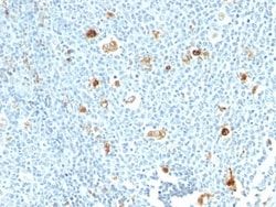
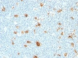
Flow Cytometry 0.5 - 1 ug/million cells in 0.1 ml, Immunohistochemistry-Paraffin 0.5 - 1 ug/ml, Immunohistochemistry-Frozen (Negative), Immunofluorescence 0.5 - 1.0 ug/ml
Gene Alias
CAGBMigration inhibitory factor-related protein 14, calgranulin B, calgranulin-B, Calprotectin L1H subunit, CFAGMRP-14,60B8AG, CGLB, L1AG, LIAG, MAC387, MIF, MRP14Leukocyte L1 complex heavy chain, NIF, P14, protein S100-A9, S100 calcium binding protein A9, S100 calcium binding protein A9 (calgranulin B), S100 calcium-binding protein A9, S100 calcium-binding protein A9 (calgranulin B)
Host Species
Mouse
Molecular Weight of Antigen
14 kDa
Quantity
0.1 mg
Research Discipline
Cancer, Immunology, Innate Immunity, Signal Transduction, Tyrosine Kinases
Gene ID (Entrez)
6280
Target Species
Human
Form
Purified
Applications
Flow Cytometry, Immunohistochemistry (Paraffin), Immunohistochemistry (Frozen), Immunofluorescence
Clone
CPT/1028
Conjugate
Unconjugated
Formulation
1.0mM PBS and 0.05% BSA with 0.05% Sodium Azide
Gene Symbols
S100A9
Immunogen
Recombinant human Calprotectin protein
Purification Method
Protein A or G purified
Regulatory Status
RUO
Primary or Secondary
Primary
Test Specificity
Recognizes the L1 or Calprotectin molecule, an intra-cytoplasmic antigen comprising of a 12kDa alpha chain and a 14kDa beta chain. Calprotectin comprises 60% of the cytoplasmic protein fraction of circulating polymorphonuclear granulocytes and is also found in monocytes, macrophages and ileal tissue eosinophils. Peripheral blood monocytes carry the antigen extra- and intracellularly, neutrophils only intracellularly. Calprotectin has antibacterial, antifungal, immunomodulating and antiproliferative effects. Besides this it is a potent chemotactic factor for neutrophils. Plasma concentrations are elevated in diseases associated with increased neutrophil activity, like inflammatory bowel disease. Granulocytes terminate their existence after transmigration through the intestinal wall. Therefore calprotectin is also detectable in feces. Elevated levels of calprotectin have been observed in body fluids such as plasma, saliva, gingival crevicular fluid, stools, and synovial fluid during infe
Content And Storage
Store at 4C.
Isotype
IgM κ
Related Products


Description
- Ensure accurate, reproducible results in Flow Cytometry, Immunohistochemistry (Paraffin), Immunofluorescence, Immunohistochemistry (Frozen) (Negative) S100A9 Monoclonal specifically detects S100A9 in Human samples
- It is validated for Immunohistochemistry, Immunohistochemistry-Paraffin.
Compare Similar Items
Show Difference
Antigen: S100A9
Classification: Monoclonal
Concentration: 0.2 mg/mL
Dilution: Flow Cytometry 0.5 - 1 ug/million cells in 0.1 ml, Immunohistochemistry-Paraffin 0.5 - 1 ug/ml, Immunohistochemistry-Frozen (Negative), Immunofluorescence 0.5 - 1.0 ug/ml
Gene Alias: CAGBMigration inhibitory factor-related protein 14, calgranulin B, calgranulin-B, Calprotectin L1H subunit, CFAGMRP-14,60B8AG, CGLB, L1AG, LIAG, MAC387, MIF, MRP14Leukocyte L1 complex heavy chain, NIF, P14, protein S100-A9, S100 calcium binding protein A9, S100 calcium binding protein A9 (calgranulin B), S100 calcium-binding protein A9, S100 calcium-binding protein A9 (calgranulin B)
Host Species: Mouse
Molecular Weight of Antigen: 14 kDa
Quantity: 0.1 mg
Research Discipline: Cancer, Immunology, Innate Immunity, Signal Transduction, Tyrosine Kinases
Gene ID (Entrez): 6280
Target Species: Human
Form: Purified
Applications: Flow Cytometry, Immunohistochemistry (Paraffin), Immunohistochemistry (Frozen), Immunofluorescence
Clone: CPT/1028
Conjugate: Unconjugated
Formulation: 1.0mM PBS and 0.05% BSA with 0.05% Sodium Azide
Gene Symbols: S100A9
Immunogen: Recombinant human Calprotectin protein
Purification Method: Protein A or G purified
Regulatory Status: RUO
Primary or Secondary: Primary
Test Specificity: Recognizes the L1 or Calprotectin molecule, an intra-cytoplasmic antigen comprising of a 12kDa alpha chain and a 14kDa beta chain. Calprotectin comprises 60% of the cytoplasmic protein fraction of circulating polymorphonuclear granulocytes and is also found in monocytes, macrophages and ileal tissue eosinophils. Peripheral blood monocytes carry the antigen extra- and intracellularly, neutrophils only intracellularly. Calprotectin has antibacterial, antifungal, immunomodulating and antiproliferative effects. Besides this it is a potent chemotactic factor for neutrophils. Plasma concentrations are elevated in diseases associated with increased neutrophil activity, like inflammatory bowel disease. Granulocytes terminate their existence after transmigration through the intestinal wall. Therefore calprotectin is also detectable in feces. Elevated levels of calprotectin have been observed in body fluids such as plasma, saliva, gingival crevicular fluid, stools, and synovial fluid during infe
Content And Storage: Store at 4C.
Isotype: IgM κ
Antigen: S100A9
Classification: Monoclonal
Concentration: 0.2 mg/mL
Dilution: Flow Cytometry 0.5 - 1 ug/million cells in 0.1 ml, Immunohistochemistry-Paraffin 0.5 - 1 ug/ml, Immunohistochemistry-Frozen (Negative), Immunofluorescence 0.5 - 1.0 ug/ml
Gene Alias: CAGBMigration inhibitory factor-related protein 14, calgranulin B, calgranulin-B, Calprotectin L1H subunit, CFAGMRP-14,60B8AG, CGLB, L1AG, LIAG, MAC387, MIF, MRP14Leukocyte L1 complex heavy chain, NIF, P14, protein S100-A9, S100 calcium binding protein A9, S100 calcium binding protein A9 (calgranulin B), S100 calcium-binding protein A9, S100 calcium-binding protein A9 (calgranulin B)
Host Species: Mouse
Molecular Weight of Antigen: 14 kDa
Quantity: 0.2 mg
Research Discipline: Cancer, Immunology, Innate Immunity, Signal Transduction, Tyrosine Kinases
Gene ID (Entrez): 6280
Target Species: Human
Form: Purified
Applications: Flow Cytometry, Immunohistochemistry (Paraffin), Immunohistochemistry (Frozen), Immunofluorescence
Clone: CPT/1028
Conjugate: Unconjugated
Formulation: 1.0mM PBS and 0.05% BSA with 0.05% Sodium Azide
Gene Symbols: S100A9
Immunogen: Recombinant human Calprotectin protein
Purification Method: Protein A or G purified
Regulatory Status: RUO
Primary or Secondary: Primary
Test Specificity: Recognizes the L1 or Calprotectin molecule, an intra-cytoplasmic antigen comprising of a 12kDa alpha chain and a 14kDa beta chain. Calprotectin comprises 60% of the cytoplasmic protein fraction of circulating polymorphonuclear granulocytes and is also found in monocytes, macrophages and ileal tissue eosinophils. Peripheral blood monocytes carry the antigen extra- and intracellularly, neutrophils only intracellularly. Calprotectin has antibacterial, antifungal, immunomodulating and antiproliferative effects. Besides this it is a potent chemotactic factor for neutrophils. Plasma concentrations are elevated in diseases associated with increased neutrophil activity, like inflammatory bowel disease. Granulocytes terminate their existence after transmigration through the intestinal wall. Therefore calprotectin is also detectable in feces. Elevated levels of calprotectin have been observed in body fluids such as plasma, saliva, gingival crevicular fluid, stools, and synovial fluid during infe
Content And Storage: Store at 4C.
Isotype: IgM κ
Antigen: CDC2/CDK1
Classification: Monoclonal
Concentration: 0.2 mg/mL
Dilution: Flow Cytometry 0.5 - 1 ug/million cells in 0.1 ml, Immunohistochemistry-Paraffin 2 - 4 ug/ml, Immunofluorescence 1 - 2 ug/ml
Gene Alias: CDC28A, CDC2MGC111195, cell cycle controller CDC2, Cell division control protein 2 homolog, Cell division protein kinase 1, cyclin-dependent kinase 1, DKFZp686L20222, EC 2.7.11.22, EC 2.7.11.23, G1 to S and G2 to M, p34 protein kinase, P34CDC2
Host Species: Mouse
Molecular Weight of Antigen: 34 kDa
Quantity: 0.02 mg
Research Discipline: Breast Cancer, Cancer, Cell Cycle and Replication, Core ESC Like Genes, Mitotic Regulators, Stem Cell Markers
Gene ID (Entrez): 983
Target Species: Human, Monkey, Mink, Primate, S. pombe (Negative), Drosophila (Negative), Mouse (Negative), Rat (Negative)
Form: Purified
Applications: Flow Cytometry, Immunohistochemistry (Paraffin), Immunofluorescence
Clone: CDK1/873
Conjugate: Unconjugated
Formulation: 10mM PBS and 0.05% BSA with 0.05% Sodium Azide
Gene Symbols: CDK1
Immunogen: Recombinant human CDK1 protein
Purification Method: Protein A or G purified
Regulatory Status: RUO
Primary or Secondary: Primary
Test Specificity: Recognizes a 34kDa protein, which is identified as cyclin dependent kinase 1 (cdk1) or p34cdc2 protein kinase. cdk1/ p34cdc2 plays a crucial role during cell division and is most active during mitosis. It is predominantly localized in the nucleus. It is a serine/threonine kinase, which is activated by cyclin, presumably by de-phosphorylation of tyrosine residues. Activated cdk1/ p34cdc2 performs specific functions during mitosis, including nuclear envelope breakdown and chromosome condensation.
Content And Storage: Store at 4C.
Isotype: IgG2a κ