CDC2/CDK1 Antibody (CDK1/873), Novus Biologicals™
Manufacturer: Fischer Scientific
Select a Size
| Pack Size | SKU | Availability | Price |
|---|---|---|---|
| Each of 1 | NBP24491500-Each-of-1 | In Stock | ₹ 23,852.00 |
NBP24491500 - Each of 1
In Stock
Quantity
1
Base Price: ₹ 23,852.00
GST (18%): ₹ 4,293.36
Total Price: ₹ 28,145.36
Antigen
CDC2/CDK1
Classification
Monoclonal
Concentration
0.2 mg/mL
Dilution
Flow Cytometry 0.5 - 1 ug/million cells in 0.1 ml, Immunohistochemistry-Paraffin 2 - 4 ug/ml, Immunofluorescence 1 - 2 ug/ml
Gene Alias
CDC28A, CDC2MGC111195, cell cycle controller CDC2, Cell division control protein 2 homolog, Cell division protein kinase 1, cyclin-dependent kinase 1, DKFZp686L20222, EC 2.7.11.22, EC 2.7.11.23, G1 to S and G2 to M, p34 protein kinase, P34CDC2
Host Species
Mouse
Molecular Weight of Antigen
34 kDa
Quantity
0.02 mg
Research Discipline
Breast Cancer, Cancer, Cell Cycle and Replication, Core ESC Like Genes, Mitotic Regulators, Stem Cell Markers
Gene ID (Entrez)
983
Target Species
Human, Monkey, Mink, Primate, S. pombe (Negative), Drosophila (Negative), Mouse (Negative), Rat (Negative)
Form
Purified
Applications
Flow Cytometry, Immunohistochemistry (Paraffin), Immunofluorescence
Clone
CDK1/873
Conjugate
Unconjugated
Formulation
10mM PBS and 0.05% BSA with 0.05% Sodium Azide
Gene Symbols
CDK1
Immunogen
Recombinant human CDK1 protein
Purification Method
Protein A or G purified
Regulatory Status
RUO
Primary or Secondary
Primary
Test Specificity
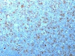
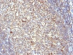
Recognizes a 34kDa protein, which is identified as cyclin dependent kinase 1 (cdk1) or p34cdc2 protein kinase. cdk1/ p34cdc2 plays a crucial role during cell division and is most active during mitosis. It is predominantly localized in the nucleus. It is a serine/threonine kinase, which is activated by cyclin, presumably by de-phosphorylation of tyrosine residues. Activated cdk1/ p34cdc2 performs specific functions during mitosis, including nuclear envelope breakdown and chromosome condensation.
Content And Storage
Store at 4C.
Isotype
IgG2a κ
Related Products


Description
- Ensure accurate, reproducible results in Flow Cytometry, Immunohistochemistry (Paraffin), Immunofluorescence CDC2/CDK1 Monoclonal specifically detects CDC2/CDK1 in Human, Bovine, Mink, Monkey, S
- pombe (Negative), Drosophila (Negative), Mouse (Negative), Rat (Negative), Xenopus (Negative), Yeast (Negative) samples
- It is validated for Western Blot, Immunohistochemistry, Immunohistochemistry-Paraffin.
Compare Similar Items
Show Difference
Antigen: CDC2/CDK1
Classification: Monoclonal
Concentration: 0.2 mg/mL
Dilution: Flow Cytometry 0.5 - 1 ug/million cells in 0.1 ml, Immunohistochemistry-Paraffin 2 - 4 ug/ml, Immunofluorescence 1 - 2 ug/ml
Gene Alias: CDC28A, CDC2MGC111195, cell cycle controller CDC2, Cell division control protein 2 homolog, Cell division protein kinase 1, cyclin-dependent kinase 1, DKFZp686L20222, EC 2.7.11.22, EC 2.7.11.23, G1 to S and G2 to M, p34 protein kinase, P34CDC2
Host Species: Mouse
Molecular Weight of Antigen: 34 kDa
Quantity: 0.02 mg
Research Discipline: Breast Cancer, Cancer, Cell Cycle and Replication, Core ESC Like Genes, Mitotic Regulators, Stem Cell Markers
Gene ID (Entrez): 983
Target Species: Human, Monkey, Mink, Primate, S. pombe (Negative), Drosophila (Negative), Mouse (Negative), Rat (Negative)
Form: Purified
Applications: Flow Cytometry, Immunohistochemistry (Paraffin), Immunofluorescence
Clone: CDK1/873
Conjugate: Unconjugated
Formulation: 10mM PBS and 0.05% BSA with 0.05% Sodium Azide
Gene Symbols: CDK1
Immunogen: Recombinant human CDK1 protein
Purification Method: Protein A or G purified
Regulatory Status: RUO
Primary or Secondary: Primary
Test Specificity: Recognizes a 34kDa protein, which is identified as cyclin dependent kinase 1 (cdk1) or p34cdc2 protein kinase. cdk1/ p34cdc2 plays a crucial role during cell division and is most active during mitosis. It is predominantly localized in the nucleus. It is a serine/threonine kinase, which is activated by cyclin, presumably by de-phosphorylation of tyrosine residues. Activated cdk1/ p34cdc2 performs specific functions during mitosis, including nuclear envelope breakdown and chromosome condensation.
Content And Storage: Store at 4C.
Isotype: IgG2a κ
Antigen: CDC2/CDK1
Classification: Monoclonal
Concentration: 0.2 mg/mL
Dilution: Flow Cytometry 0.5 - 1 ug/million cells in 0.1 ml, Immunohistochemistry-Paraffin 2 - 4 ug/ml, Immunofluorescence 1 - 2 ug/ml
Gene Alias: CDC28A, CDC2MGC111195, cell cycle controller CDC2, Cell division control protein 2 homolog, Cell division protein kinase 1, cyclin-dependent kinase 1, DKFZp686L20222, EC 2.7.11.22, EC 2.7.11.23, G1 to S and G2 to M, p34 protein kinase, P34CDC2
Host Species: Mouse
Molecular Weight of Antigen: 34 kDa
Quantity: 0.1 mg
Research Discipline: Breast Cancer, Cancer, Cell Cycle and Replication, Core ESC Like Genes, Mitotic Regulators, Stem Cell Markers
Gene ID (Entrez): 983
Target Species: Human, Monkey, Mink, Primate, S. pombe (Negative), Drosophila (Negative), Mouse (Negative), Rat (Negative)
Form: Purified
Applications: Flow Cytometry, Immunohistochemistry (Paraffin), Immunofluorescence
Clone: CDK1/873
Conjugate: Unconjugated
Formulation: 10mM PBS and 0.05% BSA with 0.05% Sodium Azide
Gene Symbols: CDK1
Immunogen: Recombinant human CDK1 protein
Purification Method: Protein A or G purified
Regulatory Status: RUO
Primary or Secondary: Primary
Test Specificity: Recognizes a 34kDa protein, which is identified as cyclin dependent kinase 1 (cdk1) or p34cdc2 protein kinase. cdk1/ p34cdc2 plays a crucial role during cell division and is most active during mitosis. It is predominantly localized in the nucleus. It is a serine/threonine kinase, which is activated by cyclin, presumably by de-phosphorylation of tyrosine residues. Activated cdk1/ p34cdc2 performs specific functions during mitosis, including nuclear envelope breakdown and chromosome condensation.
Content And Storage: Store at 4C.
Isotype: IgG2a κ
Antigen: CDC2/CDK1
Classification: Monoclonal
Concentration: 0.2 mg/mL
Dilution: Flow Cytometry 0.5 - 1 ug/million cells in 0.1 ml, Immunohistochemistry-Paraffin 2 - 4 ug/ml, Immunofluorescence 1 - 2 ug/ml
Gene Alias: CDC28A, CDC2MGC111195, cell cycle controller CDC2, Cell division control protein 2 homolog, Cell division protein kinase 1, cyclin-dependent kinase 1, DKFZp686L20222, EC 2.7.11.22, EC 2.7.11.23, G1 to S and G2 to M, p34 protein kinase, P34CDC2
Host Species: Mouse
Molecular Weight of Antigen: 34 kDa
Quantity: 0.2 mg
Research Discipline: Breast Cancer, Cancer, Cell Cycle and Replication, Core ESC Like Genes, Mitotic Regulators, Stem Cell Markers
Gene ID (Entrez): 983
Target Species: Human, Monkey, Mink, Primate, S. pombe (Negative), Drosophila (Negative), Mouse (Negative), Rat (Negative)
Form: Purified
Applications: Flow Cytometry, Immunohistochemistry (Paraffin), Immunofluorescence
Clone: CDK1/873
Conjugate: Unconjugated
Formulation: 10mM PBS and 0.05% BSA with 0.05% Sodium Azide
Gene Symbols: CDK1
Immunogen: Recombinant human CDK1 protein
Purification Method: Protein A or G purified
Regulatory Status: RUO
Primary or Secondary: Primary
Test Specificity: Recognizes a 34kDa protein, which is identified as cyclin dependent kinase 1 (cdk1) or p34cdc2 protein kinase. cdk1/ p34cdc2 plays a crucial role during cell division and is most active during mitosis. It is predominantly localized in the nucleus. It is a serine/threonine kinase, which is activated by cyclin, presumably by de-phosphorylation of tyrosine residues. Activated cdk1/ p34cdc2 performs specific functions during mitosis, including nuclear envelope breakdown and chromosome condensation.
Content And Storage: Store at 4C.
Isotype: IgG2a κ