Isocitrate Dehydrogenase 1/IDH1 Antibody (IDH1/1152), Novus Biologicals™
Manufacturer: Fischer Scientific
Select a Size
| Pack Size | SKU | Availability | Price |
|---|---|---|---|
| Each of 1 | NBP24515601-Each-of-1 | In Stock | ₹ 46,636.00 |
NBP24515601 - Each of 1
In Stock
Quantity
1
Base Price: ₹ 46,636.00
GST (18%): ₹ 8,394.48
Total Price: ₹ 55,030.48
Antigen
Isocitrate Dehydrogenase 1/IDH1
Classification
Monoclonal
Concentration
0.2mg/mL
Dilution
Western Blot 0.5 - 1.0 ug/ml, Flow Cytometry 0.5 - 1 ug/million cells in 0.1 ml, Immunohistochemistry-Paraffin 0.5 - 1.0 ug/ml, Immunofluorescence 0.5 - 1.0 ug/ml
Gene Accession No.
O75874
Gene Symbols
IDH1
Immunogen
Recombinant fragment (119 Amino acid residues around aa 280-420) of human IDH1 protein
Purification Method
Protein A or G purified
Regulatory Status
RUO
Primary or Secondary
Primary
Test Specificity
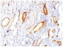
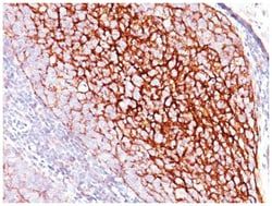
It recognizes a 45kDa protein, which is identified as isocitrate dehydrogenase (IDH1). It belongs to the isocitrate and isopropylmalate dehydrogenases family. IDH1 catalyzes the third step of the citric acid cycle, which involves the oxidative decarboxylation of isocitrate, forming ketoglutarate and CO2 in a two-step reaction. The first step involves the oxidation of isocitrate to the intermediate oxalosuccinate, while the second step involves the production of ketoglutarate. During this process, either NADH or NADPH is produced along with CO2. Recently, an inactivating mutation of IDH1 has been implicated in glioblastoma. IDH1 appears to function as a tumor suppressor that, when mutationally inactivated, contributes to tumorigenesis in part through induction of the HIF-1 pathway.
Content And Storage
Store at 4C.
Isotype
IgG1 κ
Applications
Western Blot, Flow Cytometry, Immunohistochemistry (Paraffin), Immunofluorescence
Clone
IDH1/1152
Conjugate
Unconjugated
Formulation
1.0mM PBS and 0.05% BSA with 0.05% Sodium Azide
Gene Alias
Cytosolic NADP-isocitrate dehydrogenase, EC 1.1.1.42, IDCD, IDH, IDP, IDPC, isocitrate dehydrogenase [NADP] cytoplasmic, isocitrate dehydrogenase 1 (NADP+), soluble, NADP(+)-specific ICDH, NADP-dependent isocitrate dehydrogenase, cytosolic, NADP-dependent isocitrate dehydrogenase, peroxisomal, Oxalosuccinate decarboxylase, PICD
Host Species
Mouse
Molecular Weight of Antigen
46 kDa
Quantity
0.1 mg
Research Discipline
Stem Cell Markers
Gene ID (Entrez)
3417
Target Species
Human
Form
Purified
Related Products


Description
- Ensure accurate, reproducible results in Western Blot, Flow Cytometry, Immunohistochemistry (Paraffin), Immunofluorescence Isocitrate Dehydrogenase 1/IDH1 Monoclonal specifically detects Isocitrate Dehydrogenase 1/IDH1 in Human samples
- It is validated for Immunohistochemistry, Immunohistochemistry-Paraffin.
Compare Similar Items
Show Difference
Antigen: Isocitrate Dehydrogenase 1/IDH1
Classification: Monoclonal
Concentration: 0.2mg/mL
Dilution: Western Blot 0.5 - 1.0 ug/ml, Flow Cytometry 0.5 - 1 ug/million cells in 0.1 ml, Immunohistochemistry-Paraffin 0.5 - 1.0 ug/ml, Immunofluorescence 0.5 - 1.0 ug/ml
Gene Accession No.: O75874
Gene Symbols: IDH1
Immunogen: Recombinant fragment (119 Amino acid residues around aa 280-420) of human IDH1 protein
Purification Method: Protein A or G purified
Regulatory Status: RUO
Primary or Secondary: Primary
Test Specificity: It recognizes a 45kDa protein, which is identified as isocitrate dehydrogenase (IDH1). It belongs to the isocitrate and isopropylmalate dehydrogenases family. IDH1 catalyzes the third step of the citric acid cycle, which involves the oxidative decarboxylation of isocitrate, forming ketoglutarate and CO2 in a two-step reaction. The first step involves the oxidation of isocitrate to the intermediate oxalosuccinate, while the second step involves the production of ketoglutarate. During this process, either NADH or NADPH is produced along with CO2. Recently, an inactivating mutation of IDH1 has been implicated in glioblastoma. IDH1 appears to function as a tumor suppressor that, when mutationally inactivated, contributes to tumorigenesis in part through induction of the HIF-1 pathway.
Content And Storage: Store at 4C.
Isotype: IgG1 κ
Applications: Western Blot, Flow Cytometry, Immunohistochemistry (Paraffin), Immunofluorescence
Clone: IDH1/1152
Conjugate: Unconjugated
Formulation: 1.0mM PBS and 0.05% BSA with 0.05% Sodium Azide
Gene Alias: Cytosolic NADP-isocitrate dehydrogenase, EC 1.1.1.42, IDCD, IDH, IDP, IDPC, isocitrate dehydrogenase [NADP] cytoplasmic, isocitrate dehydrogenase 1 (NADP+), soluble, NADP(+)-specific ICDH, NADP-dependent isocitrate dehydrogenase, cytosolic, NADP-dependent isocitrate dehydrogenase, peroxisomal, Oxalosuccinate decarboxylase, PICD
Host Species: Mouse
Molecular Weight of Antigen: 46 kDa
Quantity: 0.1 mg
Research Discipline: Stem Cell Markers
Gene ID (Entrez): 3417
Target Species: Human
Form: Purified
Antigen: Isocitrate Dehydrogenase 1/IDH1
Classification: Monoclonal
Concentration: 0.2mg/mL
Dilution: Western Blot 0.5 - 1.0 ug/ml, Flow Cytometry 0.5 - 1 ug/million cells in 0.1 ml, Immunohistochemistry-Paraffin 0.5 - 1.0 ug/ml, Immunofluorescence 0.5 - 1.0 ug/ml
Gene Accession No.: O75874
Gene Symbols: IDH1
Immunogen: Recombinant fragment (119 Amino acid residues around aa 280-420) of human IDH1 protein
Purification Method: Protein A or G purified
Regulatory Status: RUO
Primary or Secondary: Primary
Test Specificity: It recognizes a 45kDa protein, which is identified as isocitrate dehydrogenase (IDH1). It belongs to the isocitrate and isopropylmalate dehydrogenases family. IDH1 catalyzes the third step of the citric acid cycle, which involves the oxidative decarboxylation of isocitrate, forming ketoglutarate and CO2 in a two-step reaction. The first step involves the oxidation of isocitrate to the intermediate oxalosuccinate, while the second step involves the production of ketoglutarate. During this process, either NADH or NADPH is produced along with CO2. Recently, an inactivating mutation of IDH1 has been implicated in glioblastoma. IDH1 appears to function as a tumor suppressor that, when mutationally inactivated, contributes to tumorigenesis in part through induction of the HIF-1 pathway.
Content And Storage: Store at 4C.
Isotype: IgG1 κ
Applications: Western Blot, Flow Cytometry, Immunohistochemistry (Paraffin), Immunofluorescence
Clone: IDH1/1152
Conjugate: Unconjugated
Formulation: 1.0mM PBS and 0.05% BSA with 0.05% Sodium Azide
Gene Alias: Cytosolic NADP-isocitrate dehydrogenase, EC 1.1.1.42, IDCD, IDH, IDP, IDPC, isocitrate dehydrogenase [NADP] cytoplasmic, isocitrate dehydrogenase 1 (NADP+), soluble, NADP(+)-specific ICDH, NADP-dependent isocitrate dehydrogenase, cytosolic, NADP-dependent isocitrate dehydrogenase, peroxisomal, Oxalosuccinate decarboxylase, PICD
Host Species: Mouse
Molecular Weight of Antigen: 46 kDa
Quantity: 0.2 mg
Research Discipline: Stem Cell Markers
Gene ID (Entrez): 3417
Target Species: Human
Form: Purified
Antigen: Cadherin-16
Classification: Monoclonal
Concentration: 0.2mg/mL
Dilution: Flow Cytometry 0.5 - 1 ug/million cells in 0.1 ml, Immunohistochemistry-Paraffin 0.5 - 1.0 ug/ml, Immunofluorescence 1 - 2 ug/ml
Gene Accession No.: O75309
Gene Symbols: CDH16
Immunogen: Recombinant human CDH16 protein
Purification Method: Protein A or G purified
Regulatory Status: RUO
Primary or Secondary: Primary
Test Specificity: This MAb recognizes a protein of 130kDa, identified as Ksp-cadherin. Cadherins form a superfamily of related glycoproteins that mediate calcium-dependent cell adhesion and transmit signals from the extracellular matrix to the cytoplasm. Cadherins have been implicated in embryogenesis, tissue morphogenesis, tissue structure maintenance, cell polarization, neoplastic invasiveness and metastasis, and membrane transport. It is suggested that Ksp-cadherin is a marker for terminal differentiation of the basolateral membranes of renal tubular epithelial cells. Within the kidney, Ksp-Cadherin is found exclusively in the basolateral membrane of renal tubular epithelial cells and collecting duct cells, and not in glomeruli, renal interstitial cells, or blood vessels.Ksp-Cadherin has been suggested to distinguish Chromophobe Renal-Cell Carcinoma from Oncocytoma.
Content And Storage: Store at 4C.
Isotype: IgG1 κ
Applications: Flow Cytometry, Immunohistochemistry (Paraffin), Immunofluorescence
Clone: CDH16/1071
Conjugate: Unconjugated
Formulation: 10mM PBS and 0.05% BSA with 0.05% Sodium Azide
Gene Alias: cadherin 16, KSP-cadherin, cadherin-16, Kidney-specific cadherin, KSP-cadherin
Host Species: Mouse
Molecular Weight of Antigen: 130 kDa
Quantity: 0.02 mg
Research Discipline: __
Gene ID (Entrez): 1014
Target Species: Human, Mouse, Rat, Canine, Rabbit
Form: Purified