Mouse anti-TLR4, Clone: TLR4/230, Novus Biologicals™
Manufacturer: Fischer Scientific
The price for this product is unavailable. Please request a quote
Antigen
TLR4
Classification
Monoclonal
Concentration
0.2 mg/mL
Dilution
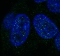
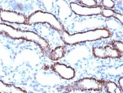
Flow Cytometry 0.5 - 1 ug/million cells in 0.1 ml, Immunohistochemistry-Paraffin 0.5 - 1.0 ug/ml, Immunofluorescence 1 - 2 ug/ml
Gene Accession No.
O00206
Gene Symbols
TLR4
Immunogen
Recombinant human TLR4 protein
Quantity
0.2 mg
Research Discipline
Adaptive Immunity, Cytokine Research, Immunology, Innate Immunity, Signal Transduction, Toll Like Receptors
Gene ID (Entrez)
7099
Target Species
Human, Rat, Porcine, Canine, Guinea Pig, Monkey
Form
Purified
Applications
Flow Cytometry, Immunohistochemistry (Paraffin), Immunofluorescence
Clone
TLR4/230
Conjugate
Unconjugated
Formulation
10mM PBS and 0.05% BSA with 0.05% Sodium Azide
Gene Alias
ARMD10, CD284, CD284 antigen, hTollhomolog of Drosophila toll, TOLL, toll-like receptor 4
Host Species
Mouse
Purification Method
Protein A or G purified
Regulatory Status
RUO
Primary or Secondary
Primary
Test Specificity
This MAb reacts with human Toll-like receptor 2 (TLR4). It is a member of the Toll-like receptor (TLR) family, which plays a fundamental role in pathogen recognition and activation of innate immunity. TLRs are highly conserved from Drosophila to humans and share structural and functional similarities. They recognize pathogen-associated molecular patterns that are expressed on infectious agents, and mediate the production of cytokines necessary for the development of effective immunity. The various TLRs exhibit different patterns of expression. This receptor has been implicated in signal transduction events induced by lipopolysaccharide (LPS) found in most gram-negative bacteria. Mutations in this gene have been associated with differences in LPS responsiveness. Multiple transcript variants encoding different isoforms have been found for this gene.
Content And Storage
Store at 4C.
Isotype
IgG2a κ
Related Products


Description
- Ensure accurate, reproducible results in Flow Cytometry, Immunohistochemistry (Paraffin), Immunofluorescence TLR4 Monoclonal specifically detects TLR4 in Human, Rat, Porcine, Canine, Guinea Pig, Monkey samples
- It is validated for Functional.
Compare Similar Items
Show Difference
Antigen: TLR4
Classification: Monoclonal
Concentration: 0.2 mg/mL
Dilution: Flow Cytometry 0.5 - 1 ug/million cells in 0.1 ml, Immunohistochemistry-Paraffin 0.5 - 1.0 ug/ml, Immunofluorescence 1 - 2 ug/ml
Gene Accession No.: O00206
Gene Symbols: TLR4
Immunogen: Recombinant human TLR4 protein
Quantity: 0.2 mg
Research Discipline: Adaptive Immunity, Cytokine Research, Immunology, Innate Immunity, Signal Transduction, Toll Like Receptors
Gene ID (Entrez): 7099
Target Species: Human, Rat, Porcine, Canine, Guinea Pig, Monkey
Form: Purified
Applications: Flow Cytometry, Immunohistochemistry (Paraffin), Immunofluorescence
Clone: TLR4/230
Conjugate: Unconjugated
Formulation: 10mM PBS and 0.05% BSA with 0.05% Sodium Azide
Gene Alias: ARMD10, CD284, CD284 antigen, hTollhomolog of Drosophila toll, TOLL, toll-like receptor 4
Host Species: Mouse
Purification Method: Protein A or G purified
Regulatory Status: RUO
Primary or Secondary: Primary
Test Specificity: This MAb reacts with human Toll-like receptor 2 (TLR4). It is a member of the Toll-like receptor (TLR) family, which plays a fundamental role in pathogen recognition and activation of innate immunity. TLRs are highly conserved from Drosophila to humans and share structural and functional similarities. They recognize pathogen-associated molecular patterns that are expressed on infectious agents, and mediate the production of cytokines necessary for the development of effective immunity. The various TLRs exhibit different patterns of expression. This receptor has been implicated in signal transduction events induced by lipopolysaccharide (LPS) found in most gram-negative bacteria. Mutations in this gene have been associated with differences in LPS responsiveness. Multiple transcript variants encoding different isoforms have been found for this gene.
Content And Storage: Store at 4C.
Isotype: IgG2a κ
Antigen: Histone H1
Classification: Monoclonal
Concentration: 0.2mg/mL
Dilution: Flow Cytometry 0.5 - 1 ug/million cells in 0.1 ml, Immunohistochemistry-Paraffin 0.5 - 1.0 ug/ml, Immunofluorescence 0.5 - 1.0 ug/ml
Gene Accession No.: P07305
Gene Symbols: H1
Immunogen: Recombinant full-length human Histone H1 protein
Quantity: 0.02 mg
Research Discipline: __
Gene ID (Entrez): 3024
Target Species: Human, Mouse, Rat
Form: Purified
Applications: Flow Cytometry, Immunohistochemistry (Paraffin), Immunofluorescence
Clone: HH1/957
Conjugate: Unconjugated
Formulation: 1.0mM PBS and 0.05% BSA with 0.05% Sodium Azide
Gene Alias: H1 histone family, member 1, H1.1, H1A, H1F1HIST1, histone 1, H1a, histone cluster 1, H1a, histone H1.1, MGC126642, MGC138345
Host Species: Mouse
Purification Method: Protein A or G purified
Regulatory Status: RUO
Primary or Secondary: Primary
Test Specificity: Eukaryotic histones are basic and water-soluble nuclear proteins that form hetero-octameric nucleosome particles by wrapping 146 base pairs of DNA in a left-handed super-helical turn sequentially to form chromosomal fiber. Two molecules of each of the four core histones (H2A, H2B, H3, and H4) form the octamer; formed of two H2A-H2B dimers and two H3-H4 dimers, forming two nearly symmetrical halves by tertiary structure. Over 80% of nucleosomes contain the linker Histone H1, derived from an intronless gene that interacts with linker DNA between nucleosomes and mediates compaction into higher order chromatin. Histones are subject to posttranslational modification by enzymes primarily on their N-terminal tails, but also in their globular domains. Such modifications include methylation, citrullination, acetylation, phosphorylation, sumoylation, ubiquitination and ADP-ribosylation.
Content And Storage: Store at 4C.
Isotype: IgG2a κ
Antigen: Histone H1
Classification: Monoclonal
Concentration: 0.2mg/mL
Dilution: Flow Cytometry 0.5 - 1 ug/million cells in 0.1 ml, Immunohistochemistry-Paraffin 0.5 - 1.0 ug/ml, Immunofluorescence 0.5 - 1.0 ug/ml
Gene Accession No.: P07305
Gene Symbols: H1
Immunogen: Recombinant full-length human Histone H1 protein
Quantity: 0.1 mg
Research Discipline: __
Gene ID (Entrez): 3024
Target Species: Human, Mouse, Rat
Form: Purified
Applications: Flow Cytometry, Immunohistochemistry (Paraffin), Immunofluorescence
Clone: HH1/957
Conjugate: Unconjugated
Formulation: 1.0mM PBS and 0.05% BSA with 0.05% Sodium Azide
Gene Alias: H1 histone family, member 1, H1.1, H1A, H1F1HIST1, histone 1, H1a, histone cluster 1, H1a, histone H1.1, MGC126642, MGC138345
Host Species: Mouse
Purification Method: Protein A or G purified
Regulatory Status: RUO
Primary or Secondary: Primary
Test Specificity: Eukaryotic histones are basic and water-soluble nuclear proteins that form hetero-octameric nucleosome particles by wrapping 146 base pairs of DNA in a left-handed super-helical turn sequentially to form chromosomal fiber. Two molecules of each of the four core histones (H2A, H2B, H3, and H4) form the octamer; formed of two H2A-H2B dimers and two H3-H4 dimers, forming two nearly symmetrical halves by tertiary structure. Over 80% of nucleosomes contain the linker Histone H1, derived from an intronless gene that interacts with linker DNA between nucleosomes and mediates compaction into higher order chromatin. Histones are subject to posttranslational modification by enzymes primarily on their N-terminal tails, but also in their globular domains. Such modifications include methylation, citrullination, acetylation, phosphorylation, sumoylation, ubiquitination and ADP-ribosylation.
Content And Storage: Store at 4C.
Isotype: IgG2a κ