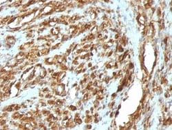
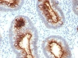
Actin (Muscle Specific) Antibody (HHF35 + MSA/953), Novus Biologicals™
Manufacturer: Fischer Scientific
Select a Size
| Pack Size | SKU | Availability | Price |
|---|---|---|---|
| Each of 1 | NBP24527500-Each-of-1 | In Stock | ₹ 23,852.00 |
NBP24527500 - Each of 1
In Stock
Quantity
1
Base Price: ₹ 23,852.00
GST (18%): ₹ 4,293.36
Total Price: ₹ 28,145.36
Antigen
pan Actin
Classification
Monoclonal
Concentration
0.2mg/mL
Dilution
Flow Cytometry 0.5 - 1 ug/million cells in 0.1 ml, Immunohistochemistry-Paraffin 0.25 - 0.5 ug/ml, Immunofluorescence 0.5 - 1.0 ug/ml
Gene Accession No.
P62736, P68032, P68133
Gene Symbols
ACTA1
Immunogen
Recombinant human actin fragment
Quantity
0.02 mg
Research Discipline
Angiogenesis, Cancer, Cell Biology, Cellular Markers, Cytoskeleton Markers, Stem Cell Markers
Gene ID (Entrez)
58
Target Species
Human, Rat, Rabbit
Form
Purified
Applications
Flow Cytometry, Immunohistochemistry (Paraffin), Immunofluorescence
Clone
HHF35 + MSA/953
Conjugate
Unconjugated
Formulation
10mM PBS and 0.05% BSA with 0.05% Sodium Azide
Gene Alias
ACTA, actin, alpha 1, skeletal muscle, alpha skeletal muscle, alpha skeletal muscle actin, alpha-actin-1, ASMA, CFTD, CFTDM, MPFD, NEM1, NEM2, NEM3
Host Species
Mouse
Purification Method
Protein A or G purified
Regulatory Status
RUO
Primary or Secondary
Primary
Test Specificity
This antibody recognizes actin of skeletal, cardiac, and smooth muscle cells. It is not reactive with other mesenchymal cells except for myoepithelium. Actin can be resolved on the basis of its isoelectric points into three distinctive components: alpha, beta, and gamma in order of increasing isoelectric point. Anti-muscle specific actin recognizes alpha and gamma isotypes of all muscle groups. Non-muscle cells such as vascular endothelial cells and connective tissues are non-reactive. Also, neoplastic cells of non-muscle-derived tissue such as carcinomas, melanomas, and lymphomas are negative.AIt stains tumors of smooth muscle (leiomyomas and leiomyosarcomas) as well as skeletal muscle (rhabdomyomas and rhabdomyosarcomas).
Content And Storage
Store at 4C.
Isotype
IgG1 κ
Related Products


Description
- Ensure accurate, reproducible results in Flow Cytometry, Immunohistochemistry (Paraffin), Immunofluorescence Actin (Muscle Specific) Monoclonal specifically detects Actin (Muscle Specific) in Human, Rat, Rabbit samples
- It is validated for Flow Cytometry, Immunohistochemistry, Immunocytochemistry/Immunofluorescence, Immunohistochemistry-Paraffin, Immunofluorescence.
Compare Similar Items
Show Difference
Antigen: pan Actin
Classification: Monoclonal
Concentration: 0.2mg/mL
Dilution: Flow Cytometry 0.5 - 1 ug/million cells in 0.1 ml, Immunohistochemistry-Paraffin 0.25 - 0.5 ug/ml, Immunofluorescence 0.5 - 1.0 ug/ml
Gene Accession No.: P62736, P68032, P68133
Gene Symbols: ACTA1
Immunogen: Recombinant human actin fragment
Quantity: 0.02 mg
Research Discipline: Angiogenesis, Cancer, Cell Biology, Cellular Markers, Cytoskeleton Markers, Stem Cell Markers
Gene ID (Entrez): 58
Target Species: Human, Rat, Rabbit
Form: Purified
Applications: Flow Cytometry, Immunohistochemistry (Paraffin), Immunofluorescence
Clone: HHF35 + MSA/953
Conjugate: Unconjugated
Formulation: 10mM PBS and 0.05% BSA with 0.05% Sodium Azide
Gene Alias: ACTA, actin, alpha 1, skeletal muscle, alpha skeletal muscle, alpha skeletal muscle actin, alpha-actin-1, ASMA, CFTD, CFTDM, MPFD, NEM1, NEM2, NEM3
Host Species: Mouse
Purification Method: Protein A or G purified
Regulatory Status: RUO
Primary or Secondary: Primary
Test Specificity: This antibody recognizes actin of skeletal, cardiac, and smooth muscle cells. It is not reactive with other mesenchymal cells except for myoepithelium. Actin can be resolved on the basis of its isoelectric points into three distinctive components: alpha, beta, and gamma in order of increasing isoelectric point. Anti-muscle specific actin recognizes alpha and gamma isotypes of all muscle groups. Non-muscle cells such as vascular endothelial cells and connective tissues are non-reactive. Also, neoplastic cells of non-muscle-derived tissue such as carcinomas, melanomas, and lymphomas are negative.AIt stains tumors of smooth muscle (leiomyomas and leiomyosarcomas) as well as skeletal muscle (rhabdomyomas and rhabdomyosarcomas).
Content And Storage: Store at 4C.
Isotype: IgG1 κ
Antigen: pan Actin
Classification: Monoclonal
Concentration: 0.2mg/mL
Dilution: Flow Cytometry 0.5 - 1 ug/million cells in 0.1 ml, Immunohistochemistry-Paraffin 0.25 - 0.5 ug/ml, Immunofluorescence 0.5 - 1.0 ug/ml
Gene Accession No.: P62736, P68032, P68133
Gene Symbols: ACTA1
Immunogen: Recombinant human actin fragment
Quantity: 0.1 mg
Research Discipline: Angiogenesis, Cancer, Cell Biology, Cellular Markers, Cytoskeleton Markers, Stem Cell Markers
Gene ID (Entrez): 58
Target Species: Human, Rat, Rabbit
Form: Purified
Applications: Flow Cytometry, Immunohistochemistry (Paraffin), Immunofluorescence
Clone: HHF35 + MSA/953
Conjugate: Unconjugated
Formulation: 10mM PBS and 0.05% BSA with 0.05% Sodium Azide
Gene Alias: ACTA, actin, alpha 1, skeletal muscle, alpha skeletal muscle, alpha skeletal muscle actin, alpha-actin-1, ASMA, CFTD, CFTDM, MPFD, NEM1, NEM2, NEM3
Host Species: Mouse
Purification Method: Protein A or G purified
Regulatory Status: RUO
Primary or Secondary: Primary
Test Specificity: This antibody recognizes actin of skeletal, cardiac, and smooth muscle cells. It is not reactive with other mesenchymal cells except for myoepithelium. Actin can be resolved on the basis of its isoelectric points into three distinctive components: alpha, beta, and gamma in order of increasing isoelectric point. Anti-muscle specific actin recognizes alpha and gamma isotypes of all muscle groups. Non-muscle cells such as vascular endothelial cells and connective tissues are non-reactive. Also, neoplastic cells of non-muscle-derived tissue such as carcinomas, melanomas, and lymphomas are negative.AIt stains tumors of smooth muscle (leiomyomas and leiomyosarcomas) as well as skeletal muscle (rhabdomyomas and rhabdomyosarcomas).
Content And Storage: Store at 4C.
Isotype: IgG1 κ
Antigen: pan Actin
Classification: Monoclonal
Concentration: 0.2mg/mL
Dilution: Flow Cytometry 0.5 - 1 ug/million cells in 0.1 ml, Immunohistochemistry-Paraffin 0.25 - 0.5 ug/ml, Immunofluorescence 0.5 - 1.0 ug/ml
Gene Accession No.: P62736, P68032, P68133
Gene Symbols: ACTA1
Immunogen: Recombinant human actin fragment
Quantity: 0.2 mg
Research Discipline: Angiogenesis, Cancer, Cell Biology, Cellular Markers, Cytoskeleton Markers, Stem Cell Markers
Gene ID (Entrez): 58
Target Species: Human, Rat, Rabbit
Form: Purified
Applications: Flow Cytometry, Immunohistochemistry (Paraffin), Immunofluorescence
Clone: HHF35 + MSA/953
Conjugate: Unconjugated
Formulation: 10mM PBS and 0.05% BSA with 0.05% Sodium Azide
Gene Alias: ACTA, actin, alpha 1, skeletal muscle, alpha skeletal muscle, alpha skeletal muscle actin, alpha-actin-1, ASMA, CFTD, CFTDM, MPFD, NEM1, NEM2, NEM3
Host Species: Mouse
Purification Method: Protein A or G purified
Regulatory Status: RUO
Primary or Secondary: Primary
Test Specificity: This antibody recognizes actin of skeletal, cardiac, and smooth muscle cells. It is not reactive with other mesenchymal cells except for myoepithelium. Actin can be resolved on the basis of its isoelectric points into three distinctive components: alpha, beta, and gamma in order of increasing isoelectric point. Anti-muscle specific actin recognizes alpha and gamma isotypes of all muscle groups. Non-muscle cells such as vascular endothelial cells and connective tissues are non-reactive. Also, neoplastic cells of non-muscle-derived tissue such as carcinomas, melanomas, and lymphomas are negative.AIt stains tumors of smooth muscle (leiomyomas and leiomyosarcomas) as well as skeletal muscle (rhabdomyomas and rhabdomyosarcomas).
Content And Storage: Store at 4C.
Isotype: IgG1 κ