B7-2/CD86 Antibody (C86/1146) - Azide and BSA Free, Novus Biologicals™
Manufacturer: Fischer Scientific
Select a Size
| Pack Size | SKU | Availability | Price |
|---|---|---|---|
| Each of 1 | NBP247778A-Each-of-1 | In Stock | ₹ 57,494.00 |
NBP247778A - Each of 1
In Stock
Quantity
1
Base Price: ₹ 57,494.00
GST (18%): ₹ 10,348.92
Total Price: ₹ 67,842.92
Antigen
B7-2/CD86
Classification
Monoclonal
Concentration
1.0 mg/mL
Dilution
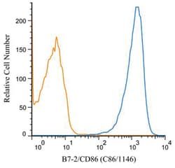
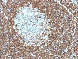
Western Blot : 0.5 - 1.0 ug/ml, Flow Cytometry : 1 ug/mL, Immunohistochemistry-Paraffin : 2 - 4 ug/ml, Flow (Cell Surface) 1:10-1:1000, Immunofluorescence : 0.5 - 1.0 ug/ml, CyTOF-ready, Knockout Validated
Gene Alias
Activation B7-2 antigen, B70, B7-2, B7-2 antigen, B-lymphocyte activation antigen B7-2, BU63, CD28 antigen ligand 2, CD28LG2B7-2 antigen), CD86 antigen, CD86 molecule, CTLA-4 counter-receptor B7.2, FUN-1, LAB72, MGC34413, T-lymphocyte activation antigen CD86
Host Species
Mouse
Molecular Weight of Antigen
70 kDa
Quantity
0.1 mg
Research Discipline
Adaptive Immunity, B Cell Development and Differentiation Markers, Cell Cycle and Replication, Dendritic Cell Markers, Diabetes Research, Immunology, Myeloid derived Suppressor Cell
Gene ID (Entrez)
942
Target Species
Human, Mouse, Rat
Form
Purified
Applications
Western Blot, Flow Cytometry, Immunohistochemistry (Paraffin), Immunofluorescence, CyTOF
Clone
C86/1146
Conjugate
Unconjugated
Formulation
PBS with No Preservative
Gene Symbols
CD86
Immunogen
Recombinant human full-length CD86 protein
Purification Method
Protein A or G purified
Regulatory Status
RUO
Primary or Secondary
Primary
Test Specificity
Recognizes a protein of 70kDa, which is identified as CD86. CD86 is a type I transmembrane glycoprotein and a member of the immunoglobulin superfamily of cell surface receptors. It is expressed at high levels on resting peripheral monocytes and dendritic cells and at very low density on resting B and T lymphocytes. CD86 expression is rapidly upregulated by B cell specific stimuli with peak expression at 18 to 42 hours after stimulation. CD86, along with CD80/B71, is an important accessory molecule in T cell co-stimulation via its interaction with CD28 and CD152/CTLA4. Since CD86 has rapid kinetics of induction, it is believed to be the major CD28 ligand expressed early in the immune response. It is also found on malignant Hodgkin and Reed Sternberg (HRS) cells in Hodgkin's disease.
Content And Storage
Store at 4C short term. Aliquot and store at -20C long term. Avoid freeze-thaw cycles.
Isotype
IgG1 κ
Related Products


Description
- B7-2/CD86 Monoclonal specifically detects B7-2/CD86 in Human, Mouse, Rat samples
- It is validated for Western Blot, Flow Cytometry, Immunohistochemistry, Immunocytochemistry/Immunofluorescence, Immunohistochemistry-Paraffin, Flow (Cell Surface), Immunofluorescence, CyTOF-ready, Knockout Validated.
Compare Similar Items
Show Difference
Antigen: B7-2/CD86
Classification: Monoclonal
Concentration: 1.0 mg/mL
Dilution: Western Blot : 0.5 - 1.0 ug/ml, Flow Cytometry : 1 ug/mL, Immunohistochemistry-Paraffin : 2 - 4 ug/ml, Flow (Cell Surface) 1:10-1:1000, Immunofluorescence : 0.5 - 1.0 ug/ml, CyTOF-ready, Knockout Validated
Gene Alias: Activation B7-2 antigen, B70, B7-2, B7-2 antigen, B-lymphocyte activation antigen B7-2, BU63, CD28 antigen ligand 2, CD28LG2B7-2 antigen), CD86 antigen, CD86 molecule, CTLA-4 counter-receptor B7.2, FUN-1, LAB72, MGC34413, T-lymphocyte activation antigen CD86
Host Species: Mouse
Molecular Weight of Antigen: 70 kDa
Quantity: 0.1 mg
Research Discipline: Adaptive Immunity, B Cell Development and Differentiation Markers, Cell Cycle and Replication, Dendritic Cell Markers, Diabetes Research, Immunology, Myeloid derived Suppressor Cell
Gene ID (Entrez): 942
Target Species: Human, Mouse, Rat
Form: Purified
Applications: Western Blot, Flow Cytometry, Immunohistochemistry (Paraffin), Immunofluorescence, CyTOF
Clone: C86/1146
Conjugate: Unconjugated
Formulation: PBS with No Preservative
Gene Symbols: CD86
Immunogen: Recombinant human full-length CD86 protein
Purification Method: Protein A or G purified
Regulatory Status: RUO
Primary or Secondary: Primary
Test Specificity: Recognizes a protein of 70kDa, which is identified as CD86. CD86 is a type I transmembrane glycoprotein and a member of the immunoglobulin superfamily of cell surface receptors. It is expressed at high levels on resting peripheral monocytes and dendritic cells and at very low density on resting B and T lymphocytes. CD86 expression is rapidly upregulated by B cell specific stimuli with peak expression at 18 to 42 hours after stimulation. CD86, along with CD80/B71, is an important accessory molecule in T cell co-stimulation via its interaction with CD28 and CD152/CTLA4. Since CD86 has rapid kinetics of induction, it is believed to be the major CD28 ligand expressed early in the immune response. It is also found on malignant Hodgkin and Reed Sternberg (HRS) cells in Hodgkin's disease.
Content And Storage: Store at 4C short term. Aliquot and store at -20C long term. Avoid freeze-thaw cycles.
Isotype: IgG1 κ
Antigen: B7-2/CD86
Classification: Monoclonal
Concentration: 1.0 mg/mL
Dilution: Western Blot : 0.5 - 1.0 ug/ml, Flow Cytometry : 1 ug/mL, Immunohistochemistry-Paraffin : 2 - 4 ug/ml, Flow (Cell Surface) 1:10-1:1000, Immunofluorescence : 0.5 - 1.0 ug/ml, CyTOF-ready, Knockout Validated
Gene Alias: Activation B7-2 antigen, B70, B7-2, B7-2 antigen, B-lymphocyte activation antigen B7-2, BU63, CD28 antigen ligand 2, CD28LG2B7-2 antigen), CD86 antigen, CD86 molecule, CTLA-4 counter-receptor B7.2, FUN-1, LAB72, MGC34413, T-lymphocyte activation antigen CD86
Host Species: Mouse
Molecular Weight of Antigen: 70 kDa
Quantity: 0.2 mg
Research Discipline: Adaptive Immunity, B Cell Development and Differentiation Markers, Cell Cycle and Replication, Dendritic Cell Markers, Diabetes Research, Immunology, Myeloid derived Suppressor Cell
Gene ID (Entrez): 942
Target Species: Human, Mouse, Rat
Form: Purified
Applications: Western Blot, Flow Cytometry, Immunohistochemistry (Paraffin), Immunofluorescence, CyTOF
Clone: C86/1146
Conjugate: Unconjugated
Formulation: PBS with No Preservative
Gene Symbols: CD86
Immunogen: Recombinant human full-length CD86 protein
Purification Method: Protein A or G purified
Regulatory Status: RUO
Primary or Secondary: Primary
Test Specificity: Recognizes a protein of 70kDa, which is identified as CD86. CD86 is a type I transmembrane glycoprotein and a member of the immunoglobulin superfamily of cell surface receptors. It is expressed at high levels on resting peripheral monocytes and dendritic cells and at very low density on resting B and T lymphocytes. CD86 expression is rapidly upregulated by B cell specific stimuli with peak expression at 18 to 42 hours after stimulation. CD86, along with CD80/B71, is an important accessory molecule in T cell co-stimulation via its interaction with CD28 and CD152/CTLA4. Since CD86 has rapid kinetics of induction, it is believed to be the major CD28 ligand expressed early in the immune response. It is also found on malignant Hodgkin and Reed Sternberg (HRS) cells in Hodgkin's disease.
Content And Storage: Store at 4C short term. Aliquot and store at -20C long term. Avoid freeze-thaw cycles.
Isotype: IgG1 κ
Antigen: PMEL17/SILV
Classification: Monoclonal
Concentration: 1.0 mg/mL
Dilution: Flow Cytometry : 0.5 - 1 ug/million cells in 0.1 ml, Immunohistochemistry-Paraffin : 0.5 - 1.0 ug/ml, Immunofluorescence : 0.5 - 1.0 ug/ml, CyTOF-ready
Gene Alias: D12S53EP1, gp100, ME20, ME20-M, melanocyte protein mel 17, Melanocyte protein Pmel 17, Melanocytes lineage-specific antigen GP100, Melanoma-associated ME20 antigen, melanosomal matrix protein17, PMEL17P100, premelanosome proteinME20M, SI, SIL, silver (mouse homolog) like, silver homolog (mouse), Silver locus protein homolog, silver, mouse, homolog of, SILVPmel17
Host Species: Mouse
Molecular Weight of Antigen: 95 kDa
Quantity: 0.1 mg
Research Discipline: __
Gene ID (Entrez): 6490
Target Species: Human
Form: Purified
Applications: Flow Cytometry, Immunohistochemistry (Paraffin), Immunofluorescence, CyTOF
Clone: PMEL/783
Conjugate: Unconjugated
Formulation: PBS with No Preservative
Gene Symbols: PMEL
Immunogen: Recombinant human SILV protein
Purification Method: Protein A or G purified
Regulatory Status: RUO
Primary or Secondary: Primary
Test Specificity: Cytotoxic T lymphocytes (CTLs) recognize melanoma-associated antigens, which belong to three main groups. These groups include tumor-associated testis-specific antigens, melanocyte differentiation antigens and mutated or aberrantly expressed antigens, which are routinely used as markers to identify melanomas based on their binding to specific monoclonal antibodies. gp100, also designated ME20-M, ME20-S and PMEL 17, is classified as a melanocyte differentiation antigen and is expressed at low levels in normal cell lines and tissues, but is upregulated in melanocytes. gp100 is a highly glycosylated protein. It is also the product of proteolytic cleavage, which results in a secreted protein
Content And Storage: Store at 4C short term. Aliquot and store at -20C long term. Avoid freeze-thaw cycles.
Isotype: IgG1 κ