Pax6 Antibody (PAX6/1166) - Azide and BSA Free, Novus Biologicals™
Manufacturer: Fischer Scientific
The price for this product is unavailable. Please request a quote
Antigen
Pax6
Classification
Monoclonal
Concentration
1.0 mg/mL
Dilution
Flow Cytometry : 0.5 - 1 ug/million cells in 0.1 ml, Immunohistochemistry-Paraffin : 0.5 - 1.0 ug/ml, Immunofluorescence : 0.5 - 1.0 ug/ml, CyTOF-ready
Gene Alias
keratitis), MGC17209, Oculorhombin, paired box 6, paired box protein Pax-6
Host Species
Mouse
Molecular Weight of Antigen
47 kDa
Quantity
0.2 mg
Research Discipline
Cellular Markers, Diabetes Research, Neuronal Stem Cell Markers, Neuronal Stem Cells, Neuroscience, Sensory Systems, Stem Cell Markers, Stem Cells, Transcription Factors and Regulators, Vision
Gene ID (Entrez)
5080
Target Species
Human
Form
Purified
Applications
Flow Cytometry, Immunohistochemistry (Paraffin), Immunofluorescence, CyTOF
Clone
PAX6/1166
Conjugate
Unconjugated
Formulation
PBS with No Preservative
Gene Symbols
PAX6
Immunogen
Recombinant fragment (N-terminus; aa 1-300) of human PAX6 protein
Purification Method
Protein A or G purified
Regulatory Status
RUO
Primary or Secondary
Primary
Test Specificity
Pax genes contain paired domains with strong homology to genes in Drosophila, which are involved in programming early development. Lesions in the Pax-6 gene account for most cases of aniridia, a congenital malformation of the eye, chiefly characterized by iris hypoplasia, which can cause blindness. Pax-6 is involved in other anterior segment malformations besides aniridia, such as Peters anomaly, a major error in the embryonic development of the eye with corneal clouding with variable iridolenticulocorneal adhesions. The Pax-6 gene encodes a transcriptional regulator that recognizes target genes through its paired-type DNA-binding domain. The paired domain is composed of two distinct DNA-binding subdomains, the amino-terminal subdomain and the carboxy-terminal subdomain, which bind respective consensus DNA sequences. The human Pax-6 gene produces two alternatively spliced isoforms that have the distinct structure of the paired domain.
Content And Storage
Store at 4C short term. Aliquot and store at -20C long term. Avoid freeze-thaw cycles.
Isotype
IgG1 κ
Related Products


Description
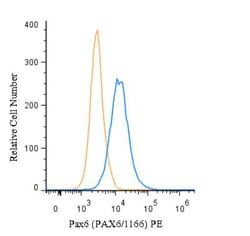
- Pax6 Monoclonal specifically detects Pax6 in Human samples
- It is validated for Flow Cytometry, Immunohistochemistry, Immunohistochemistry-Paraffin, Flow (Intracellular), CyTOF-ready, Knockout Validated.
Compare Similar Items
Show Difference
Antigen: Pax6
Classification: Monoclonal
Concentration: 1.0 mg/mL
Dilution: Flow Cytometry : 0.5 - 1 ug/million cells in 0.1 ml, Immunohistochemistry-Paraffin : 0.5 - 1.0 ug/ml, Immunofluorescence : 0.5 - 1.0 ug/ml, CyTOF-ready
Gene Alias: keratitis), MGC17209, Oculorhombin, paired box 6, paired box protein Pax-6
Host Species: Mouse
Molecular Weight of Antigen: 47 kDa
Quantity: 0.2 mg
Research Discipline: Cellular Markers, Diabetes Research, Neuronal Stem Cell Markers, Neuronal Stem Cells, Neuroscience, Sensory Systems, Stem Cell Markers, Stem Cells, Transcription Factors and Regulators, Vision
Gene ID (Entrez): 5080
Target Species: Human
Form: Purified
Applications: Flow Cytometry, Immunohistochemistry (Paraffin), Immunofluorescence, CyTOF
Clone: PAX6/1166
Conjugate: Unconjugated
Formulation: PBS with No Preservative
Gene Symbols: PAX6
Immunogen: Recombinant fragment (N-terminus; aa 1-300) of human PAX6 protein
Purification Method: Protein A or G purified
Regulatory Status: RUO
Primary or Secondary: Primary
Test Specificity: Pax genes contain paired domains with strong homology to genes in Drosophila, which are involved in programming early development. Lesions in the Pax-6 gene account for most cases of aniridia, a congenital malformation of the eye, chiefly characterized by iris hypoplasia, which can cause blindness. Pax-6 is involved in other anterior segment malformations besides aniridia, such as Peters anomaly, a major error in the embryonic development of the eye with corneal clouding with variable iridolenticulocorneal adhesions. The Pax-6 gene encodes a transcriptional regulator that recognizes target genes through its paired-type DNA-binding domain. The paired domain is composed of two distinct DNA-binding subdomains, the amino-terminal subdomain and the carboxy-terminal subdomain, which bind respective consensus DNA sequences. The human Pax-6 gene produces two alternatively spliced isoforms that have the distinct structure of the paired domain.
Content And Storage: Store at 4C short term. Aliquot and store at -20C long term. Avoid freeze-thaw cycles.
Isotype: IgG1 κ
Antigen: Moesin
Classification: Monoclonal
Concentration: 1.0 mg/mL
Dilution: Western Blot : 0.5 - 1.0 ug/ml, Flow Cytometry : 0.5 - 1 ug/million cells in 0.1 ml, Immunohistochemistry-Paraffin : 0.5 - 1.0 ug/ml, Immunofluorescence : 0.5 - 1.0 ug/ml, CyTOF-ready
Gene Alias: Membrane-organizing extension spike protein, moesin
Host Species: Mouse
Molecular Weight of Antigen: 78 kDa
Quantity: 0.1 mg
Research Discipline: Cytoskeleton Markers, Stem Cell Markers
Gene ID (Entrez): 4478
Target Species: Human, Rat (Negative)
Form: Purified
Applications: Western Blot, Flow Cytometry, Immunohistochemistry (Paraffin), Immunofluorescence, CyTOF
Clone: MSN/492
Conjugate: Unconjugated
Formulation: PBS with No Preservative
Gene Symbols: MSN
Immunogen: Recombinant full-length human Moesin protein
Purification Method: Protein A or G purified
Regulatory Status: RUO
Primary or Secondary: Primary
Test Specificity: Recognizes 78kDa moesin protein. Moesin, a member of the talin-4.1 superfamily, is a linking protein of the sub-membranous actin cytoskeleton. It is expressed in variable amounts in cells of different phenotypes such as macrophages, lymphocytes, fibroblastic, endothelial, epithelial, and neuronal cell lines but not in blood cells. The ERM proteins, ezrin, radixin, and moesin are involved in a variety of cellular functions, such as cell adhesion, migration, and the organization of cell surface structures, and are highly homologous, both in protein sequence and in functional activity, with merlin/schwannomin, a neurofibromatosis-2-associated tumor-suppressor protein. Cell lines of epithelial and mesothelial origin contain both moesin and radixin whereas cells of endothelial and lymphoid origin express moesin.
Content And Storage: Store at 4C short term. Aliquot and store at -20C long term. Avoid freeze-thaw cycles.
Isotype: IgG1 κ
Antigen: Moesin
Classification: Monoclonal
Concentration: 1.0 mg/mL
Dilution: Western Blot : 0.5 - 1.0 ug/ml, Flow Cytometry : 0.5 - 1 ug/million cells in 0.1 ml, Immunohistochemistry-Paraffin : 0.5 - 1.0 ug/ml, Immunofluorescence : 0.5 - 1.0 ug/ml, CyTOF-ready
Gene Alias: Membrane-organizing extension spike protein, moesin
Host Species: Mouse
Molecular Weight of Antigen: 78 kDa
Quantity: 0.2 mg
Research Discipline: Cytoskeleton Markers, Stem Cell Markers
Gene ID (Entrez): 4478
Target Species: Human, Rat (Negative)
Form: Purified
Applications: Western Blot, Flow Cytometry, Immunohistochemistry (Paraffin), Immunofluorescence, CyTOF
Clone: MSN/492
Conjugate: Unconjugated
Formulation: PBS with No Preservative
Gene Symbols: MSN
Immunogen: Recombinant full-length human Moesin protein
Purification Method: Protein A or G purified
Regulatory Status: RUO
Primary or Secondary: Primary
Test Specificity: Recognizes 78kDa moesin protein. Moesin, a member of the talin-4.1 superfamily, is a linking protein of the sub-membranous actin cytoskeleton. It is expressed in variable amounts in cells of different phenotypes such as macrophages, lymphocytes, fibroblastic, endothelial, epithelial, and neuronal cell lines but not in blood cells. The ERM proteins, ezrin, radixin, and moesin are involved in a variety of cellular functions, such as cell adhesion, migration, and the organization of cell surface structures, and are highly homologous, both in protein sequence and in functional activity, with merlin/schwannomin, a neurofibromatosis-2-associated tumor-suppressor protein. Cell lines of epithelial and mesothelial origin contain both moesin and radixin whereas cells of endothelial and lymphoid origin express moesin.
Content And Storage: Store at 4C short term. Aliquot and store at -20C long term. Avoid freeze-thaw cycles.
Isotype: IgG1 κ