Retinol Binding Protein RBP Antibody (RBP/872) - Azide and BSA Free, Novus Biologicals™
Manufacturer: Fischer Scientific
The price for this product is unavailable. Please request a quote
Antigen
Retinol Binding Protein RBP
Classification
Monoclonal
Concentration
1.0 mg/mL
Dilution
Immunohistochemistry-Paraffin : 1 - 2 ug/ml, Immunofluorescence : 1 - 2 ug/ml
Gene Alias
C, Cellular retinol-binding protein, CRABP-I, CRBP1CRBP-I, CRBPCellular retinol-binding protein I, CRBPI, RBPC, retinol binding protein 1, cellular, retinol-binding protein 1, retinol-binding protein 1, cellular
Host Species
Mouse
Purification Method
Protein A or G purified
Regulatory Status
RUO
Gene ID (Entrez)
5947
Target Species
Human, Mouse, Rat, Goat, Monkey, Rabbit
Form
Purified
Applications
Immunohistochemistry (Paraffin), Immunofluorescence
Clone
RBP/872
Conjugate
Unconjugated
Formulation
PBS with No Preservative
Gene Symbols
RBP1
Immunogen
Recombinant human retinol binding protein-1 (RBP1)
Quantity
0.2 mg
Primary or Secondary
Primary
Test Specificity
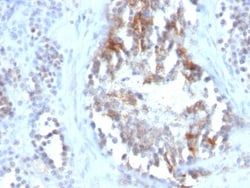
Recognizes a protein of 21kDa-25kDa, identified as retinol binding protein-1 (RBP1). This protein belongs to the lipocalin family and is the specific carrier for retinol (vitamin A alcohol) in the blood. It delivers retinol from the liver stores to the peripheral tissues. In plasma, the RBP-retinol complex interacts with transthyretin, which prevents its loss by filtration through the kidney glomeruli. A deficiency of vitamin A blocks secretion of the binding protein post-transnationally and results in defective delivery and supply to the epidermal cells.
Content And Storage
Store at 4C short term. Aliquot and store at -20C long term. Avoid freeze-thaw cycles.
Isotype
IgG1 κ
Related Products


Description
- Retinol Binding Protein RBP Monoclonal specifically detects Retinol Binding Protein RBP in Human, Mouse, Rat, Goat, Monkey, Rabbit samples
- It is validated for Immunohistochemistry, Immunohistochemistry-Paraffin.
Compare Similar Items
Show Difference
Antigen: Retinol Binding Protein RBP
Classification: Monoclonal
Concentration: 1.0 mg/mL
Dilution: Immunohistochemistry-Paraffin : 1 - 2 ug/ml, Immunofluorescence : 1 - 2 ug/ml
Gene Alias: C, Cellular retinol-binding protein, CRABP-I, CRBP1CRBP-I, CRBPCellular retinol-binding protein I, CRBPI, RBPC, retinol binding protein 1, cellular, retinol-binding protein 1, retinol-binding protein 1, cellular
Host Species: Mouse
Purification Method: Protein A or G purified
Regulatory Status: RUO
Gene ID (Entrez): 5947
Target Species: Human, Mouse, Rat, Goat, Monkey, Rabbit
Form: Purified
Applications: Immunohistochemistry (Paraffin), Immunofluorescence
Clone: RBP/872
Conjugate: Unconjugated
Formulation: PBS with No Preservative
Gene Symbols: RBP1
Immunogen: Recombinant human retinol binding protein-1 (RBP1)
Quantity: 0.2 mg
Primary or Secondary: Primary
Test Specificity: Recognizes a protein of 21kDa-25kDa, identified as retinol binding protein-1 (RBP1). This protein belongs to the lipocalin family and is the specific carrier for retinol (vitamin A alcohol) in the blood. It delivers retinol from the liver stores to the peripheral tissues. In plasma, the RBP-retinol complex interacts with transthyretin, which prevents its loss by filtration through the kidney glomeruli. A deficiency of vitamin A blocks secretion of the binding protein post-transnationally and results in defective delivery and supply to the epidermal cells.
Content And Storage: Store at 4C short term. Aliquot and store at -20C long term. Avoid freeze-thaw cycles.
Isotype: IgG1 κ
Antigen: Fibronectin
Classification: Monoclonal
Concentration: 1.0 mg/mL
Dilution: Flow Cytometry : 0.5 - 1 ug/million cells in 0.1 ml, Immunohistochemistry-Paraffin : 1 - 2 ug/ml, Immunofluorescence : 0.5 - 1.0 ug/ml, CyTOF-ready
Gene Alias: __
Host Species: Mouse
Purification Method: Protein A or G purified
Regulatory Status: RUO
Gene ID (Entrez): 2335
Target Species: Human
Form: Purified
Applications: Flow Cytometry, Immunohistochemistry (Paraffin), Immunofluorescence, CyTOF
Clone: 568
Conjugate: Unconjugated
Formulation: PBS with No Preservative
Gene Symbols: FN1
Immunogen: High molecular weight proteins secreted by cultivated human fibroblasts
Quantity: 0.1 mg
Primary or Secondary: Primary
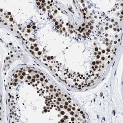
Test Specificity: Epitope of this MAb is located in the 8th type III repeat in the cell-binding region of fibronectin. Fibronectins are disulfide-linked, dimeric glycoproteins of ∼440kDa. They possess at least four binding sites for collagen, glycosaminoglycans, transglutaminase, and a cell surface receptor. Fibronectins are involved in cell adhesion, tissue organization, and wound healing. Fibronectins are present in basement membranes, interstitial connective tissue matrix, and blood. Cellular fibronectin is widely distributed in the stroma of many malignant tumors. This MAb is excellent for staining of formalin-fixed, paraffin-embedded tissues.
Content And Storage: Store at 4C short term. Aliquot and store at -20C long term. Avoid freeze-thaw cycles.
Isotype: IgG1 κ
Antigen: Fibronectin
Classification: Monoclonal
Concentration: 1.0 mg/mL
Dilution: Flow Cytometry : 0.5 - 1 ug/million cells in 0.1 ml, Immunohistochemistry-Paraffin : 1 - 2 ug/ml, Immunofluorescence : 0.5 - 1.0 ug/ml, CyTOF-ready
Gene Alias: __
Host Species: Mouse
Purification Method: Protein A or G purified
Regulatory Status: RUO
Gene ID (Entrez): 2335
Target Species: Human
Form: Purified
Applications: Flow Cytometry, Immunohistochemistry (Paraffin), Immunofluorescence, CyTOF
Clone: 568
Conjugate: Unconjugated
Formulation: PBS with No Preservative
Gene Symbols: FN1
Immunogen: High molecular weight proteins secreted by cultivated human fibroblasts
Quantity: 0.2 mg
Primary or Secondary: Primary
Test Specificity: Epitope of this MAb is located in the 8th type III repeat in the cell-binding region of fibronectin. Fibronectins are disulfide-linked, dimeric glycoproteins of ∼440kDa. They possess at least four binding sites for collagen, glycosaminoglycans, transglutaminase, and a cell surface receptor. Fibronectins are involved in cell adhesion, tissue organization, and wound healing. Fibronectins are present in basement membranes, interstitial connective tissue matrix, and blood. Cellular fibronectin is widely distributed in the stroma of many malignant tumors. This MAb is excellent for staining of formalin-fixed, paraffin-embedded tissues.
Content And Storage: Store at 4C short term. Aliquot and store at -20C long term. Avoid freeze-thaw cycles.
Isotype: IgG1 κ