SHBG Antibody (SPM605), Novus Biologicals™
Manufacturer: Fischer Scientific
The price for this product is unavailable. Please request a quote
Antigen
SHBG
Classification
Monoclonal
Concentration
0.2mg/mL
Dilution
Flow Cytometry 0.5 - 1 ug/million cells in 0.1 ml, Immunohistochemistry-Paraffin 1 - 2 ug/ml, Immunofluorescence 0.5 - 1.0 ug/ml
Gene Alias
ABPTestosterone-estrogen-binding globulin, androgen binding protein, MGC126834, MGC138391, SBP, sex hormone-binding globulin, Sex steroid-binding protein, TeBG, Testis-specific androgen-binding protein, testosterone-binding beta-globulin, Testosterone-estradiol-binding globulin
Host Species
Mouse
Molecular Weight of Antigen
45 kDa
Quantity
0.2 mg
Research Discipline
Cancer, Vision
Gene ID (Entrez)
6462
Target Species
Human
Form
Purified
Applications
Flow Cytometry, Immunohistochemistry (Paraffin), Immunofluorescence
Clone
SPM605
Conjugate
Unconjugated
Formulation
10mM PBS and 0.05% BSA with 0.05% Sodium Azide
Gene Symbols
SHBG
Immunogen
Recombinant full-length human SHBG protein
Purification Method
Protein A or G purified
Regulatory Status
RUO
Primary or Secondary
Primary
Test Specificity
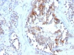
Recognizes a protein of 45kDa, identified as SHBG. It functions as an androgen transport protein, but may also be involved in receptor-mediated processes. Each dimer binds one molecule of steroid. It is specific for 5-alpha-dihydrotestosterone, testosterone, and 17-beta-estradiol. SHBG regulates the plasma metabolic clearance rate of steroid hormones by controlling their plasma concentration. In testis, it is synthesized by the Sertoli cells, secreted into the lumen of the seminiferous tubule and transported to the epididymis.
Content And Storage
Store at 4C.
Isotype
IgG1
Related Products


Description
- Ensure accurate, reproducible results in Flow Cytometry, Immunohistochemistry (Paraffin), Immunofluorescence SHBG Monoclonal specifically detects SHBG in Human samples
- It is validated for Flow Cytometry, Immunohistochemistry, Immunocytochemistry/Immunofluorescence, Immunohistochemistry-Paraffin, Immunofluorescence.
Compare Similar Items
Show Difference
Antigen: SHBG
Classification: Monoclonal
Concentration: 0.2mg/mL
Dilution: Flow Cytometry 0.5 - 1 ug/million cells in 0.1 ml, Immunohistochemistry-Paraffin 1 - 2 ug/ml, Immunofluorescence 0.5 - 1.0 ug/ml
Gene Alias: ABPTestosterone-estrogen-binding globulin, androgen binding protein, MGC126834, MGC138391, SBP, sex hormone-binding globulin, Sex steroid-binding protein, TeBG, Testis-specific androgen-binding protein, testosterone-binding beta-globulin, Testosterone-estradiol-binding globulin
Host Species: Mouse
Molecular Weight of Antigen: 45 kDa
Quantity: 0.2 mg
Research Discipline: Cancer, Vision
Gene ID (Entrez): 6462
Target Species: Human
Form: Purified
Applications: Flow Cytometry, Immunohistochemistry (Paraffin), Immunofluorescence
Clone: SPM605
Conjugate: Unconjugated
Formulation: 10mM PBS and 0.05% BSA with 0.05% Sodium Azide
Gene Symbols: SHBG
Immunogen: Recombinant full-length human SHBG protein
Purification Method: Protein A or G purified
Regulatory Status: RUO
Primary or Secondary: Primary
Test Specificity: Recognizes a protein of 45kDa, identified as SHBG. It functions as an androgen transport protein, but may also be involved in receptor-mediated processes. Each dimer binds one molecule of steroid. It is specific for 5-alpha-dihydrotestosterone, testosterone, and 17-beta-estradiol. SHBG regulates the plasma metabolic clearance rate of steroid hormones by controlling their plasma concentration. In testis, it is synthesized by the Sertoli cells, secreted into the lumen of the seminiferous tubule and transported to the epididymis.
Content And Storage: Store at 4C.
Isotype: IgG1
Antigen: Von Willebrand Factor
Classification: Monoclonal
Concentration: 0.2 mg/mL
Dilution: Western Blot 0.5 - 1.0 ug/ml, Flow Cytometry 0.5 - 1 ug/million cells in 0.1 ml, Immunoprecipitation 0.5 - 1 ug/500 ug Protein Lysate, Immunohistochemistry-Paraffin 0.5 - 1.0 ug/ml, Immunofluorescence 0.5 - 1.0 ug/ml
Gene Alias: coagulation factor VIII VWF, F8, F8VWF, von Willebrand factor, VWD, vWF
Host Species: Mouse
Molecular Weight of Antigen: 250 kDa
Quantity: 0.02 mg
Research Discipline: Cancer
Gene ID (Entrez): 7450
Target Species: Human
Form: Purified
Applications: Western Blot, Flow Cytometry, Immunoprecipitation, Immunohistochemistry (Paraffin), Immunofluorescence
Clone: IIIE2.34
Conjugate: Unconjugated
Formulation: 10mM PBS and 0.05% BSA with 0.05% Sodium Azide
Gene Symbols: VWF
Immunogen: Recombinant human vWF fragment spanning aa 845-949
Purification Method: Protein A or G purified
Regulatory Status: RUO
Primary or Secondary: Primary
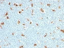
Test Specificity: Von Willebrand Factor (vWF) is a multimeric glycoprotein that is found in endothelial cells, plasma and platelets. It acts as a carrier protein for Factor VIII and promotes platelet adhesion and aggregation. vWF undergoes a variety of posttranslational modifications that influence the affinity and availability for Factor VIII, including cleavage of the propeptide and formation of N-terminal disulfide bonds. This antibody helps to establish the endothelial nature of some lesions of disputed histogenesis, e.g. Kaposi's sarcoma and cardiac myxoma. It is widely used for differentiating vascular lesions from those of other tissue differentiation within a panel of other vascular markers although not all tumors of endothelial differentiation contain this antigen.
Content And Storage: Store at 4C.
Isotype: IgG1 κ
Antigen: Von Willebrand Factor
Classification: Monoclonal
Concentration: 0.2 mg/mL
Dilution: Western Blot 0.5 - 1.0 ug/ml, Flow Cytometry 0.5 - 1 ug/million cells in 0.1 ml, Immunoprecipitation 0.5 - 1 ug/500 ug Protein Lysate, Immunohistochemistry-Paraffin 0.5 - 1.0 ug/ml, Immunofluorescence 0.5 - 1.0 ug/ml
Gene Alias: coagulation factor VIII VWF, F8, F8VWF, von Willebrand factor, VWD, vWF
Host Species: Mouse
Molecular Weight of Antigen: 250 kDa
Quantity: 0.1 mg
Research Discipline: Cancer
Gene ID (Entrez): 7450
Target Species: Human
Form: Purified
Applications: Western Blot, Flow Cytometry, Immunoprecipitation, Immunohistochemistry (Paraffin), Immunofluorescence
Clone: IIIE2.34
Conjugate: Unconjugated
Formulation: 10mM PBS and 0.05% BSA with 0.05% Sodium Azide
Gene Symbols: VWF
Immunogen: Recombinant human vWF fragment spanning aa 845-949
Purification Method: Protein A or G purified
Regulatory Status: RUO
Primary or Secondary: Primary
Test Specificity: Von Willebrand Factor (vWF) is a multimeric glycoprotein that is found in endothelial cells, plasma and platelets. It acts as a carrier protein for Factor VIII and promotes platelet adhesion and aggregation. vWF undergoes a variety of posttranslational modifications that influence the affinity and availability for Factor VIII, including cleavage of the propeptide and formation of N-terminal disulfide bonds. This antibody helps to establish the endothelial nature of some lesions of disputed histogenesis, e.g. Kaposi's sarcoma and cardiac myxoma. It is widely used for differentiating vascular lesions from those of other tissue differentiation within a panel of other vascular markers although not all tumors of endothelial differentiation contain this antigen.
Content And Storage: Store at 4C.
Isotype: IgG1 κ