Arginase 1/ARG1/liver Arginase Antibody (ARG1/1125) - IHC-Prediluted, Novus Biologicals™
Manufacturer: Novus Biologicals
Select a Size
| Pack Size | SKU | Availability | Price |
|---|---|---|---|
| Each of 1 | NBP248229-Each-of-1 | In Stock | ₹ 46,636.00 |
NBP248229 - Each of 1
In Stock
Quantity
1
Base Price: ₹ 46,636.00
GST (18%): ₹ 8,394.48
Total Price: ₹ 55,030.48
Antigen
Arginase 1/ARG1/liver Arginase
Classification
Monoclonal
Conjugate
Unconjugated
Formulation
10mM PBS and 0.05% BSA with 0.05% Sodium Azide
Gene Symbols
ARG1
Immunogen
Recombinant human Arginase 1/ARG1/liver Arginase protein fragment (around aa11-97) (exact sequence is proprietary) (Uniprot: P05089)
Purification Method
Protein A or G purified
Regulatory Status
RUO
Primary or Secondary
Primary
Test Specificity
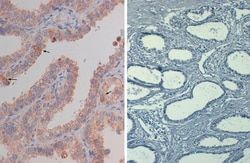
Recognizes a protein of 35-38kDa, which is identified as Arginase 1 (ARG1). Arginase is a manganese metallo-enzyme that catalyzes the hydrolysis of arginine to generate ornithine and urea. Arginase I and II are isoenzymes which differ in subcellular localization, regulation, and possibly function. Arginase I is a cytosolic enzyme, which is expressed mainly in the liver as part of the urea cycle, whereas arginase II is a mitochondrial protein found in a variety of tissues. Antibody to ARG-1 labels hepatocytes in normal tissues and granulocytes in peripheral blood. ARG-1 is a sensitive and specific marker for identification of hepatocellular carcinoma.
Content And Storage
Store at 4C.
Isotype
IgG3 κ
Applications
Immunohistochemistry (Paraffin)
Clone
ARG1/1125
Dilution
Immunohistochemistry-Paraffin
Gene Alias
arginase 1, arginase, liver, arginase-1, EC 3.5.3.1, Liver-type arginase, Type I arginase
Host Species
Mouse
Molecular Weight of Antigen
36.5 kDa
Quantity
7 mL
Research Discipline
Cancer, Cellular Markers, Chromatin Research, Lipid and Metabolism, Myeloid derived Suppressor Cell
Gene ID (Entrez)
383
Target Species
Human
Form
Purified
Related Products


Description
- Arginase 1/ARG1/liver Arginase Monoclonal specifically detects Arginase 1/ARG1/liver Arginase in Human samples
- It is validated for Immunohistochemistry, Immunohistochemistry-Paraffin.
Compare Similar Items
Show Difference
Antigen: Arginase 1/ARG1/liver Arginase
Classification: Monoclonal
Conjugate: Unconjugated
Formulation: 10mM PBS and 0.05% BSA with 0.05% Sodium Azide
Gene Symbols: ARG1
Immunogen: Recombinant human Arginase 1/ARG1/liver Arginase protein fragment (around aa11-97) (exact sequence is proprietary) (Uniprot: P05089)
Purification Method: Protein A or G purified
Regulatory Status: RUO
Primary or Secondary: Primary
Test Specificity: Recognizes a protein of 35-38kDa, which is identified as Arginase 1 (ARG1). Arginase is a manganese metallo-enzyme that catalyzes the hydrolysis of arginine to generate ornithine and urea. Arginase I and II are isoenzymes which differ in subcellular localization, regulation, and possibly function. Arginase I is a cytosolic enzyme, which is expressed mainly in the liver as part of the urea cycle, whereas arginase II is a mitochondrial protein found in a variety of tissues. Antibody to ARG-1 labels hepatocytes in normal tissues and granulocytes in peripheral blood. ARG-1 is a sensitive and specific marker for identification of hepatocellular carcinoma.
Content And Storage: Store at 4C.
Isotype: IgG3 κ
Applications: Immunohistochemistry (Paraffin)
Clone: ARG1/1125
Dilution: Immunohistochemistry-Paraffin
Gene Alias: arginase 1, arginase, liver, arginase-1, EC 3.5.3.1, Liver-type arginase, Type I arginase
Host Species: Mouse
Molecular Weight of Antigen: 36.5 kDa
Quantity: 7 mL
Research Discipline: Cancer, Cellular Markers, Chromatin Research, Lipid and Metabolism, Myeloid derived Suppressor Cell
Gene ID (Entrez): 383
Target Species: Human
Form: Purified
Antigen: Arginase 1/ARG1/liver Arginase
Classification: Monoclonal
Conjugate: Unconjugated
Formulation: 10mM PBS and 0.05% BSA with 0.05% Sodium Azide
Gene Symbols: ARG1
Immunogen: Recombinant fragment (around aa11-97) of human Arginase 1/ARG1/liver Arginase protein(exact sequence is proprietary) (Uniprot: P05089)
Purification Method: Protein A or G purified
Regulatory Status: RUO
Primary or Secondary: Primary
Test Specificity: Recognizes a protein of 35-38kDa, which is identified as Arginase 1 (ARG1). Arginase is a manganese metallo-enzyme that catalyzes the hydrolysis of arginine to generate ornithine and urea. Arginase I and II are isoenzymes, which differ in subcellular localization, regulation, and possibly function. Arginase I is a cytosolic enzyme, which is expressed mainly in the liver as part of the urea cycle, whereas arginase II is a mitochondrial protein found in a variety of tissues. Antibody to ARG-1 labels hepatocytes in normal tissues and granulocytes in peripheral blood. ARG-1 is a sensitive and specific marker for identification of hepatocellular carcinoma.
Content And Storage: Store at 4C.
Isotype: IgG3 κ
Applications: Immunohistochemistry (Paraffin)
Clone: ARG1/1126
Dilution: Immunohistochemistry-Paraffin
Gene Alias: arginase 1, arginase, liver, arginase-1, EC 3.5.3.1, Liver-type arginase, Type I arginase
Host Species: Mouse
Molecular Weight of Antigen: 36.5 kDa
Quantity: 7 mL
Research Discipline: Cancer, Cellular Markers, Chromatin Research, Lipid and Metabolism, Myeloid derived Suppressor Cell
Gene ID (Entrez): 383
Target Species: Human
Form: Purified
Antigen: Melanoma Associated Antigen (PNL2)
Classification: Monoclonal
Conjugate: Unconjugated
Formulation: 10mM PBS and 0.05% BSA with 0.05% Sodium Azide
Gene Symbols: __
Immunogen: Melanocyte antigen
Purification Method: Protein A or G purified
Regulatory Status: RUO
Primary or Secondary: Primary
Test Specificity: Anti-PNL2 is a novel monoclonal antibody, which has recently been introduced as an immunohistochemical reagent to stain melanocytes and tumors derived therefrom. The antigen recognized by PNL2 is different from Melan A and gp100. Its epitope is not destroyed by digestion with neuraminidase i.e. its epitope id not glycosylated. Anti-PNL2 may be most useful because of its high sensitivity for metastatic melanoma (87%), as opposed to 76% for anti-HMB45 and 82% for anti-MART-1. Anti-PNL2 labels intra-epidermal nevi while the dermal component of compound nevi are largely non-reactive with anti-PNL2. Antibodies against PNL2, MART-1 (Melan A) and HMB45 stain most clear cell sarcoma cells and a few cells in angio-myolipomas and lymphangioleiomyomatosis. Anti-PNL2 is a useful antibody for the identification of melanomas and clear cell sarcomas. Differential diagnosis is aided by the results from a panel of antibodies, including antibodies against HMB45, MART-1, tyrosinase, and MiTF.
Content And Storage: Store at 4C.
Isotype: IgG1 κ
Applications: Immunohistochemistry (Paraffin)
Clone: PNL2
Dilution: Immunohistochemistry-Paraffin
Gene Alias: __
Host Species: Mouse
Molecular Weight of Antigen: __
Quantity: 7 mL
Research Discipline: __
Gene ID (Entrez): __
Target Species: Human, Mouse, Canine
Form: Purified