Myoglobin Antibody, Novus Biologicals™
Manufacturer: Novus Biologicals
Select a Size
| Pack Size | SKU | Availability | Price |
|---|---|---|---|
| Each of 1 | NBP254897-Each-of-1 | In Stock | ₹ 57,983.50 |
NBP254897 - Each of 1
In Stock
Quantity
1
Base Price: ₹ 57,983.50
GST (18%): ₹ 10,437.03
Total Price: ₹ 68,420.53
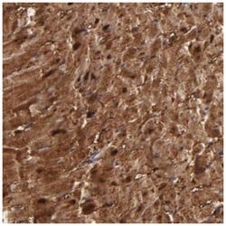
Antigen
Myoglobin
Classification
Polyclonal
Dilution
Immunohistochemistry-Paraffin 1:200 - 1:500
Gene Alias
MGC13548, myoglobin, PVALB
Host Species
Rabbit
Purification Method
Affinity Purified
Regulatory Status
RUO
Primary or Secondary
Primary
Test Specificity
Specificity of antibody verified on a Protein Array containing target protein plus 383 other non-specific proteins.
Content And Storage
Store at 4C short term. Aliquot and store at -20C long term. Avoid freeze-thaw cycles.
Applications
Immunohistochemistry (Paraffin)
Conjugate
Unconjugated
Formulation
PBS (pH 7.2) and 40% Glycerol with 0.02% Sodium Azide
Gene Symbols
MB
Immunogen
This antibody was developed against Recombinant Protein corresponding to amino acids:DGEWQLVLNVWGKVEADIPGHGQEVLIRLFKGHPETLEKFDKFKHLKSEDEMKASEDLKKHGATVLTALGGILKKKGHHEAEIKPLAQSHATKHKIPVKYLEFISECIIQVLQSKHPGDFGADA
Quantity
0.1 mL
Research Discipline
Cancer, Cardiovascular Biology
Gene ID (Entrez)
4151
Target Species
Human
Isotype
IgG
Description
- Myoglobin Polyclonal specifically detects Myoglobin in Human samples
- It is validated for Immunohistochemistry, Immunohistochemistry-Paraffin.
Compare Similar Items
Show Difference
Antigen: Myoglobin
Classification: Polyclonal
Dilution: Immunohistochemistry-Paraffin 1:200 - 1:500
Gene Alias: MGC13548, myoglobin, PVALB
Host Species: Rabbit
Purification Method: Affinity Purified
Regulatory Status: RUO
Primary or Secondary: Primary
Test Specificity: Specificity of antibody verified on a Protein Array containing target protein plus 383 other non-specific proteins.
Content And Storage: Store at 4C short term. Aliquot and store at -20C long term. Avoid freeze-thaw cycles.
Applications: Immunohistochemistry (Paraffin)
Conjugate: Unconjugated
Formulation: PBS (pH 7.2) and 40% Glycerol with 0.02% Sodium Azide
Gene Symbols: MB
Immunogen: This antibody was developed against Recombinant Protein corresponding to amino acids:DGEWQLVLNVWGKVEADIPGHGQEVLIRLFKGHPETLEKFDKFKHLKSEDEMKASEDLKKHGATVLTALGGILKKKGHHEAEIKPLAQSHATKHKIPVKYLEFISECIIQVLQSKHPGDFGADA
Quantity: 0.1 mL
Research Discipline: Cancer, Cardiovascular Biology
Gene ID (Entrez): 4151
Target Species: Human
Isotype: IgG
Antigen: SPTLC3
Classification: Polyclonal
Dilution: Western Blot 0.4 ug/ml, Immunocytochemistry/Immunofluorescence
Gene Alias: C20orf38, Chromosome 20 Open Reading Frame 38, dJ718P11, EC 2.3.1.50, hLCB2b, LCB 3, LCB2B, LCB3, Long Chain Base Biosynthesis Protein 2b, Long Chain Base Biosynthesis Protein 3, Serine Palmitoyltransferase 3, Serine Palmitoyltransferase, Long Chain Base Subunit 2-Like (Aminotransferase 2), Serine Palmitoyltransferase, Long Chain Base Subunit 3, Serine-Palmitoyl-CoA Transferase 3, SPT 3, SPT3, SPTLC2L
Host Species: Rabbit
Purification Method: Affinity Purified
Regulatory Status: RUO
Primary or Secondary: Primary
Test Specificity: Specificity of antibody verified on a Protein Array containing target protein plus 383 other non-specific proteins.
Content And Storage: Store at 4C short term. Aliquot and store at -20C long term. Avoid freeze-thaw cycles.
Applications: Western Blot, Immunocytochemistry, Immunofluorescence
Conjugate: Unconjugated
Formulation: 40% glycerol and PBS (pH 7.2) with 0.02% Sodium Azide
Gene Symbols: SPTLC3
Immunogen: Recombinant Protein Epitope Signature Tag (PrEST) antigen sequence: MANPGGGAVCNGKLHNHKKQSNGSQSRNCTKNGIVKEAQQNGKPHFYDKLIVESFEEA
Quantity: 100 μL
Research Discipline: __
Gene ID (Entrez): 55304
Target Species: Human
Isotype: IgG

Antigen: CD7
Classification: Monoclonal
Dilution: Flow Cytometry 0.5 - 1ug/million cells in 0.1 ml, Immunofluorescence 0.5 - 1.0 ug/ml
Gene Alias: CD7 antigen, CD7 antigen (p41), CD7 molecule, GP40T-cell surface antigen Leu-9, LEU-9, T-cell antigen CD7, T-cell leukemia antigen, Tp40, TP41p41 protein
Host Species: Mouse
Purification Method: Protein A or G purified
Regulatory Status: RUO
Primary or Secondary: Primary
Test Specificity: Recognizes a protein of 40kDa, identified as CD7, a member of the immunoglobulin gene superfamily. Its N-terminal amino acids 1-107 are highly homologous to Ig kappa-L chains whereas the carboxyl-terminal region of the extracellular domain is proline-rich and has been postulated to form a stalk from which the Ig domain projects. CD7 is expressed on the majority of immature and mature T-lymphocytes, and T cell leukemia. It is also found on natural killer cells, a small subpopulation of normal B cells and on malignant B cells. Cross-linking surface CD7 positively modulates T cell and NK cell activity as measured by calcium fluxes, expression of adhesion molecules, cytokine secretion and proliferation. CD7 associates directly with phosphoinositol 3'-kinase. CD7 ligation induces production of D-3 phosphoinositides and tyrosine phosphorylation.
Content And Storage: Store at 4°C.
Applications: Flow Cytometry, Immunofluorescence
Conjugate: Unconjugated
Formulation: 10mM PBS with No Preservative
Gene Symbols: CD7
Immunogen: Human T cells
Quantity: 0.1 mg
Research Discipline: Cytokine Research, Signal Transduction
Gene ID (Entrez): 924
Target Species: Human
Isotype: IgG1 κ