Factor XIIIa Mouse anti-Human, Clone: F13A1/1448, Novus Biologicals™
Manufacturer: Fischer Scientific
Select a Size
| Pack Size | SKU | Availability | Price |
|---|---|---|---|
| Each of 1 | NBP259604X-Each-of-1 | In Stock | ₹ 46,814.00 |
NBP259604X - Each of 1
In Stock
Quantity
1
Base Price: ₹ 46,814.00
GST (18%): ₹ 8,426.52
Total Price: ₹ 55,240.52
Antigen
Factor XIIIa
Classification
Monoclonal
Concentration
0.2 mg/mL
Dilution
Western Blot 0.5-1.0 ug/ml, Flow Cytometry 0.5-1 ug/million cells, ELISA 2-4 ug/ml, Immunocytochemistry/Immunofluorescence 0.5-1 ug/ml, Immunohistochemistry-Paraffin 1-2 ug/ml, SDS-Page, Protein Array 1:100-1:2000
Gene Alias
bA525O21.1 (coagulation factor XIII, A1 polypeptide), coagulation factor XIII A chain, coagulation factor XIII, A1 polypeptide, Coagulation factor XIIIa, EC 2.3.2.13, F13Acoagulation factor XIII, A polypeptide, factor XIIIa, fibrin stabilizing factor, A subunit, fibrinoligase, FSF, A subunit, Protein-glutamine gamma-glutamyltransferase A chain, TGase, Transglutaminase A chain, transglutaminase. plasma
Host Species
Mouse
Molecular Weight of Antigen
83 kDa
Quantity
100 μg
Research Discipline
Apoptosis, Cancer, Cell Biology
Gene ID (Entrez)
2162
Target Species
Human
Form
Purified
Applications
Western Blot, Flow Cytometry, ELISA, Immunocytochemistry, Immunofluorescence, Immunohistochemistry (Paraffin)
Clone
F13A1/1448
Conjugate
Unconjugated
Formulation
10mM PBS with 0.05% BSA with 0.05% Sodium Azide
Gene Symbols
F13A1
Immunogen
Recombinant fragment of human Factor XIIIa protein (aa46-181) (exact sequence is proprietary)
Purification Method
Protein A or G purified
Regulatory Status
RUO
Primary or Secondary
Primary
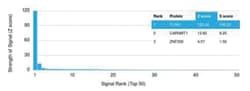
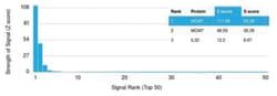
Test Specificity
The specificity of this monoclonal antibody to its intended target was validated by HuProtTM Array, containing more than 19,000, full-length human proteins. It recognizes a protein of 83kDa, which is identified as Factor XIIIa. It has been identified in platelets, megakaryocytes, and fibroblast-like mesenchymal or histiocytic cells in the placenta, uterus, and prostate, monocytes and macrophages and dermal dendritic cells. Anti-factor XIIIa has been found to be useful in differentiating between dermatofibroma (almost all cases are positive), dermatofibrosarcoma protuberans ( -/+) and desmoplastic malignant melanoma (-). Anti-factor XIIIa positivity is also seen in capillary hemagioblastoma, hemangioendothelioma, hemangiopericytoma, xanthogranuloma, xanthoma, hepatocellular carcinoma, glomus tumor, and meningioma.
Content And Storage
Store at 4C short term. Aliquot and store at -20C long term. Avoid freeze-thaw cycles.
Isotype
IgG2b κ
Related Products
Description
- Factor XIIIa Monoclonal specifically detects Factor XIIIa in Human samples
- It is validated for Western Blot, Flow Cytometry, ELISA, Immunohistochemistry, Immunocytochemistry/Immunofluorescence, Immunohistochemistry-Paraffin, Protein Array.
Compare Similar Items
Show Difference
Antigen: Factor XIIIa
Classification: Monoclonal
Concentration: 0.2 mg/mL
Dilution: Western Blot 0.5-1.0 ug/ml, Flow Cytometry 0.5-1 ug/million cells, ELISA 2-4 ug/ml, Immunocytochemistry/Immunofluorescence 0.5-1 ug/ml, Immunohistochemistry-Paraffin 1-2 ug/ml, SDS-Page, Protein Array 1:100-1:2000
Gene Alias: bA525O21.1 (coagulation factor XIII, A1 polypeptide), coagulation factor XIII A chain, coagulation factor XIII, A1 polypeptide, Coagulation factor XIIIa, EC 2.3.2.13, F13Acoagulation factor XIII, A polypeptide, factor XIIIa, fibrin stabilizing factor, A subunit, fibrinoligase, FSF, A subunit, Protein-glutamine gamma-glutamyltransferase A chain, TGase, Transglutaminase A chain, transglutaminase. plasma
Host Species: Mouse
Molecular Weight of Antigen: 83 kDa
Quantity: 100 μg
Research Discipline: Apoptosis, Cancer, Cell Biology
Gene ID (Entrez): 2162
Target Species: Human
Form: Purified
Applications: Western Blot, Flow Cytometry, ELISA, Immunocytochemistry, Immunofluorescence, Immunohistochemistry (Paraffin)
Clone: F13A1/1448
Conjugate: Unconjugated
Formulation: 10mM PBS with 0.05% BSA with 0.05% Sodium Azide
Gene Symbols: F13A1
Immunogen: Recombinant fragment of human Factor XIIIa protein (aa46-181) (exact sequence is proprietary)
Purification Method: Protein A or G purified
Regulatory Status: RUO
Primary or Secondary: Primary
Test Specificity: The specificity of this monoclonal antibody to its intended target was validated by HuProtTM Array, containing more than 19,000, full-length human proteins. It recognizes a protein of 83kDa, which is identified as Factor XIIIa. It has been identified in platelets, megakaryocytes, and fibroblast-like mesenchymal or histiocytic cells in the placenta, uterus, and prostate, monocytes and macrophages and dermal dendritic cells. Anti-factor XIIIa has been found to be useful in differentiating between dermatofibroma (almost all cases are positive), dermatofibrosarcoma protuberans ( -/+) and desmoplastic malignant melanoma (-). Anti-factor XIIIa positivity is also seen in capillary hemagioblastoma, hemangioendothelioma, hemangiopericytoma, xanthogranuloma, xanthoma, hepatocellular carcinoma, glomus tumor, and meningioma.
Content And Storage: Store at 4C short term. Aliquot and store at -20C long term. Avoid freeze-thaw cycles.
Isotype: IgG2b κ

Antigen: Factor XIIIa
Classification: Monoclonal
Concentration: __
Dilution: Western Blot, Flow Cytometry, ELISA, Immunohistochemistry, Immunocytochemistry/Immunofluorescence, Immunohistochemistry-Paraffin
Gene Alias: bA525O21.1 (coagulation factor XIII, A1 polypeptide), coagulation factor XIII A chain, coagulation factor XIII, A1 polypeptide, Coagulation factor XIIIa, EC 2.3.2.13, F13Acoagulation factor XIII, A polypeptide, factor XIIIa, fibrin stabilizing factor, A subunit, fibrinoligase, FSF, A subunit, Protein-glutamine gamma-glutamyltransferase A chain, TGase, Transglutaminase A chain, transglutaminase. plasma
Host Species: Mouse
Molecular Weight of Antigen: __
Quantity: 100 μL
Research Discipline: Apoptosis, Cancer, Cell Biology
Gene ID (Entrez): 2162
Target Species: Human
Form: Purified
Applications: Western Blot, Flow Cytometry, ELISA, Immunohistochemistry, Immunocytochemistry, Immunofluorescence, Immunohistochemistry (Paraffin)
Clone: F13A1/1448
Conjugate: Alexa Fluor 488
Formulation: 50mM Sodium Borate with 0.05% Sodium Azide
Gene Symbols: F13A1
Immunogen: Recombinant fragment of human Factor XIIIa protein (aa46-181) (exact sequence is proprietary) (Uniprot: P00488)
Purification Method: Protein A or G purified
Regulatory Status: RUO
Primary or Secondary: Primary
Test Specificity: The specificity of this monoclonal antibody to its intended target was validated by HuProtTM Array, containing more than 19,000, full-length human proteins. It recognizes a protein of 83kDa, which is identified as Factor XIIIa. It has been identified in platelets, megakaryocytes, and fibroblast-like mesenchymal or histiocytic cells in the placenta, uterus, and prostate, monocytes and macrophages and dermal dendritic cells. Anti-factor XIIIa has been found to be useful in differentiating between dermatofibroma (almost all cases are positive), dermatofibrosarcoma protuberans (-/+) and desmoplastic malignant melanoma (-). Anti-factor XIIIa positivity is also seen in capillary hemagioblastoma, hemangioendothelioma, hemangiopericytoma, xanthogranuloma, xanthoma, hepatocellular carcinoma, glomus tumor, and meningioma.
Content And Storage: Store at 4C in the dark.
Isotype: IgG2b κ
Antigen: Factor XIIIa
Classification: Monoclonal
Concentration: __
Dilution: Western Blot, Flow Cytometry, ELISA, Immunohistochemistry, Immunocytochemistry/Immunofluorescence, Immunohistochemistry-Paraffin
Gene Alias: bA525O21.1 (coagulation factor XIII, A1 polypeptide), coagulation factor XIII A chain, coagulation factor XIII, A1 polypeptide, Coagulation factor XIIIa, EC 2.3.2.13, F13Acoagulation factor XIII, A polypeptide, factor XIIIa, fibrin stabilizing factor, A subunit, fibrinoligase, FSF, A subunit, Protein-glutamine gamma-glutamyltransferase A chain, TGase, Transglutaminase A chain, transglutaminase. plasma
Host Species: Mouse
Molecular Weight of Antigen: __
Quantity: 100 μL
Research Discipline: Apoptosis, Cancer, Cell Biology
Gene ID (Entrez): 2162
Target Species: Human
Form: Purified
Applications: Western Blot, Flow Cytometry, ELISA, Immunohistochemistry, Immunocytochemistry, Immunofluorescence, Immunohistochemistry (Paraffin)
Clone: F13A1/1448
Conjugate: Alexa Fluor 405
Formulation: 50mM Sodium Borate with 0.05% Sodium Azide
Gene Symbols: F13A1
Immunogen: Recombinant fragment of human Factor XIIIa protein (aa46-181) (exact sequence is proprietary) (Uniprot: P00488)
Purification Method: Protein A or G purified
Regulatory Status: RUO
Primary or Secondary: Primary
Test Specificity: The specificity of this monoclonal antibody to its intended target was validated by HuProtTM Array, containing more than 19,000, full-length human proteins. It recognizes a protein of 83kDa, which is identified as Factor XIIIa. It has been identified in platelets, megakaryocytes, and fibroblast-like mesenchymal or histiocytic cells in the placenta, uterus, and prostate, monocytes and macrophages and dermal dendritic cells. Anti-factor XIIIa has been found to be useful in differentiating between dermatofibroma (almost all cases are positive), dermatofibrosarcoma protuberans (-/+) and desmoplastic malignant melanoma (-). Anti-factor XIIIa positivity is also seen in capillary hemagioblastoma, hemangioendothelioma, hemangiopericytoma, xanthogranuloma, xanthoma, hepatocellular carcinoma, glomus tumor, and meningioma.
Content And Storage: Store at 4C in the dark.
Isotype: IgG2b κ